{"title":"竖脊肌阻滞:急性阑尾炎疼痛管理的新方法。","authors":"Jonathan Brewer, Holly Conger, Robert Rash","doi":"10.1186/s13089-022-00281-7","DOIUrl":null,"url":null,"abstract":"<p><strong>Background: </strong>Acute abdominal pain is one of the most common complaints that patients present with in the emergency room and has long been a challenge to effectively manage without relying on opioid analgesia. The use of ultrasound-guided peripheral nerve blocks (UGRA) represents a new frontier in multimodal pain control regimens in the acute setting. An erector spinae plane (ESP) block is believed to mediate pain relief in multiple dermatomes through blockage of both visceral and somatic nerves. Analgesia provided by a single injection can help keep a patient comfortable for hours without breakthrough pain and the subsequent need for frequent redosing of opioid pain medication. To this date, there is very limited evidence of an ESP block in the utilization of acute appendicitis in the emergency department.</p><p><strong>Case report: </strong>This case report presents a 26-year-old female with a past medical history of polycystic ovarian syndrome (PCOS) and a tubal ligation that presented with 7/10 right lower quadrant abdominal pain that began 1 h prior to arrival. She stated that she felt like this was similar to her PCOS exacerbations in the past. During her evaluation, she underwent a computed tomography (CT) scan of her abdomen and pelvis that was remarkable for acute, uncomplicated appendicitis. She was given 4 mg of morphine for her pain with little response, so the offer was made for an erector spinae block that the patient elected to receive. After being consented both for the procedure and for research, she received a right-sided erector spinae block with 20 mL's of 0.2% ropivacaine (2 mg/mL) at the L1 vertebral level. After approximately 15 min, she stated that she had a reduction in her pain from a 6/10 to a 1/10 that persisted throughout the rest of her stay in the emergency department.</p>","PeriodicalId":36911,"journal":{"name":"Ultrasound Journal","volume":" ","pages":"30"},"PeriodicalIF":3.4000,"publicationDate":"2022-07-26","publicationTypes":"Journal Article","fieldsOfStudy":null,"isOpenAccess":false,"openAccessPdf":"https://www.ncbi.nlm.nih.gov/pmc/articles/PMC9325929/pdf/","citationCount":"0","resultStr":"{\"title\":\"The erector spinae block: a novel approach to pain management in acute appendicitis.\",\"authors\":\"Jonathan Brewer, Holly Conger, Robert Rash\",\"doi\":\"10.1186/s13089-022-00281-7\",\"DOIUrl\":null,\"url\":null,\"abstract\":\"<p><strong>Background: </strong>Acute abdominal pain is one of the most common complaints that patients present with in the emergency room and has long been a challenge to effectively manage without relying on opioid analgesia. The use of ultrasound-guided peripheral nerve blocks (UGRA) represents a new frontier in multimodal pain control regimens in the acute setting. An erector spinae plane (ESP) block is believed to mediate pain relief in multiple dermatomes through blockage of both visceral and somatic nerves. Analgesia provided by a single injection can help keep a patient comfortable for hours without breakthrough pain and the subsequent need for frequent redosing of opioid pain medication. To this date, there is very limited evidence of an ESP block in the utilization of acute appendicitis in the emergency department.</p><p><strong>Case report: </strong>This case report presents a 26-year-old female with a past medical history of polycystic ovarian syndrome (PCOS) and a tubal ligation that presented with 7/10 right lower quadrant abdominal pain that began 1 h prior to arrival. She stated that she felt like this was similar to her PCOS exacerbations in the past. During her evaluation, she underwent a computed tomography (CT) scan of her abdomen and pelvis that was remarkable for acute, uncomplicated appendicitis. She was given 4 mg of morphine for her pain with little response, so the offer was made for an erector spinae block that the patient elected to receive. After being consented both for the procedure and for research, she received a right-sided erector spinae block with 20 mL's of 0.2% ropivacaine (2 mg/mL) at the L1 vertebral level. After approximately 15 min, she stated that she had a reduction in her pain from a 6/10 to a 1/10 that persisted throughout the rest of her stay in the emergency department.</p>\",\"PeriodicalId\":36911,\"journal\":{\"name\":\"Ultrasound Journal\",\"volume\":\" \",\"pages\":\"30\"},\"PeriodicalIF\":3.4000,\"publicationDate\":\"2022-07-26\",\"publicationTypes\":\"Journal Article\",\"fieldsOfStudy\":null,\"isOpenAccess\":false,\"openAccessPdf\":\"https://www.ncbi.nlm.nih.gov/pmc/articles/PMC9325929/pdf/\",\"citationCount\":\"0\",\"resultStr\":null,\"platform\":\"Semanticscholar\",\"paperid\":null,\"PeriodicalName\":\"Ultrasound Journal\",\"FirstCategoryId\":\"1085\",\"ListUrlMain\":\"https://doi.org/10.1186/s13089-022-00281-7\",\"RegionNum\":0,\"RegionCategory\":null,\"ArticlePicture\":[],\"TitleCN\":null,\"AbstractTextCN\":null,\"PMCID\":null,\"EPubDate\":\"\",\"PubModel\":\"\",\"JCR\":\"Q2\",\"JCRName\":\"Medicine\",\"Score\":null,\"Total\":0}","platform":"Semanticscholar","paperid":null,"PeriodicalName":"Ultrasound Journal","FirstCategoryId":"1085","ListUrlMain":"https://doi.org/10.1186/s13089-022-00281-7","RegionNum":0,"RegionCategory":null,"ArticlePicture":[],"TitleCN":null,"AbstractTextCN":null,"PMCID":null,"EPubDate":"","PubModel":"","JCR":"Q2","JCRName":"Medicine","Score":null,"Total":0}
The erector spinae block: a novel approach to pain management in acute appendicitis.
Background: Acute abdominal pain is one of the most common complaints that patients present with in the emergency room and has long been a challenge to effectively manage without relying on opioid analgesia. The use of ultrasound-guided peripheral nerve blocks (UGRA) represents a new frontier in multimodal pain control regimens in the acute setting. An erector spinae plane (ESP) block is believed to mediate pain relief in multiple dermatomes through blockage of both visceral and somatic nerves. Analgesia provided by a single injection can help keep a patient comfortable for hours without breakthrough pain and the subsequent need for frequent redosing of opioid pain medication. To this date, there is very limited evidence of an ESP block in the utilization of acute appendicitis in the emergency department.
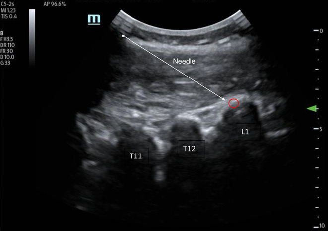
Case report: This case report presents a 26-year-old female with a past medical history of polycystic ovarian syndrome (PCOS) and a tubal ligation that presented with 7/10 right lower quadrant abdominal pain that began 1 h prior to arrival. She stated that she felt like this was similar to her PCOS exacerbations in the past. During her evaluation, she underwent a computed tomography (CT) scan of her abdomen and pelvis that was remarkable for acute, uncomplicated appendicitis. She was given 4 mg of morphine for her pain with little response, so the offer was made for an erector spinae block that the patient elected to receive. After being consented both for the procedure and for research, she received a right-sided erector spinae block with 20 mL's of 0.2% ropivacaine (2 mg/mL) at the L1 vertebral level. After approximately 15 min, she stated that she had a reduction in her pain from a 6/10 to a 1/10 that persisted throughout the rest of her stay in the emergency department.


 分享
分享
 求助内容:
求助内容: 应助结果提醒方式:
 扫码关注我们
