{"title":"有症状的巨大肝囊肿经皮引流及硬化治疗后迟发性囊内出血1例报告。","authors":"Koji Mikami, Hiroshi Yukimoto","doi":"10.22575/interventionalradiology.2021-0005","DOIUrl":null,"url":null,"abstract":"<p><p>Herein, we have reported a rare case of intracystic hemorrhage due to rupture of a right hepatic artery pseudoaneurysm in a 76-year-old female patient who underwent drainage and 3% polidocanol sclerotherapy for a symptomatic giant hepatic cyst. One month after sclerotherapy, the patient presented to the emergency room with acute and severe abdominal pain. Non-contrast T1-weighted magnetic resonance imaging findings showed high hepatic cyst fluid signal intensity and abdominal arteriography findings revealed a right hepatic artery pseudoaneurysm surrounding the hepatic cystic wall. Therefore, the patient was diagnosed with intracystic hemorrhage due to a ruptured pseudoaneurysm. Embolization, using a detachable coil, was successful. Interventional radiologists should be aware of potential vascular injuries during drainage and sclerotherapy for giant hepatic cysts.</p>","PeriodicalId":73503,"journal":{"name":"Interventional radiology (Higashimatsuyama-shi (Japan)","volume":"6 2","pages":"61-64"},"PeriodicalIF":0.8000,"publicationDate":"2021-07-01","publicationTypes":"Journal Article","fieldsOfStudy":null,"isOpenAccess":false,"openAccessPdf":"https://ftp.ncbi.nlm.nih.gov/pub/pmc/oa_pdf/82/f0/2432-0935-6-2-0061.PMC9327434.pdf","citationCount":"0","resultStr":"{\"title\":\"Delayed Intracystic Hemorrhage after Percutaneous Drainage and Sclerotherapy for a Symptomatic Giant Hepatic Cyst: A Case Report.\",\"authors\":\"Koji Mikami, Hiroshi Yukimoto\",\"doi\":\"10.22575/interventionalradiology.2021-0005\",\"DOIUrl\":null,\"url\":null,\"abstract\":\"<p><p>Herein, we have reported a rare case of intracystic hemorrhage due to rupture of a right hepatic artery pseudoaneurysm in a 76-year-old female patient who underwent drainage and 3% polidocanol sclerotherapy for a symptomatic giant hepatic cyst. One month after sclerotherapy, the patient presented to the emergency room with acute and severe abdominal pain. Non-contrast T1-weighted magnetic resonance imaging findings showed high hepatic cyst fluid signal intensity and abdominal arteriography findings revealed a right hepatic artery pseudoaneurysm surrounding the hepatic cystic wall. Therefore, the patient was diagnosed with intracystic hemorrhage due to a ruptured pseudoaneurysm. Embolization, using a detachable coil, was successful. Interventional radiologists should be aware of potential vascular injuries during drainage and sclerotherapy for giant hepatic cysts.</p>\",\"PeriodicalId\":73503,\"journal\":{\"name\":\"Interventional radiology (Higashimatsuyama-shi (Japan)\",\"volume\":\"6 2\",\"pages\":\"61-64\"},\"PeriodicalIF\":0.8000,\"publicationDate\":\"2021-07-01\",\"publicationTypes\":\"Journal Article\",\"fieldsOfStudy\":null,\"isOpenAccess\":false,\"openAccessPdf\":\"https://ftp.ncbi.nlm.nih.gov/pub/pmc/oa_pdf/82/f0/2432-0935-6-2-0061.PMC9327434.pdf\",\"citationCount\":\"0\",\"resultStr\":null,\"platform\":\"Semanticscholar\",\"paperid\":null,\"PeriodicalName\":\"Interventional radiology (Higashimatsuyama-shi (Japan)\",\"FirstCategoryId\":\"1085\",\"ListUrlMain\":\"https://doi.org/10.22575/interventionalradiology.2021-0005\",\"RegionNum\":0,\"RegionCategory\":null,\"ArticlePicture\":[],\"TitleCN\":null,\"AbstractTextCN\":null,\"PMCID\":null,\"EPubDate\":\"\",\"PubModel\":\"\",\"JCR\":\"\",\"JCRName\":\"\",\"Score\":null,\"Total\":0}","platform":"Semanticscholar","paperid":null,"PeriodicalName":"Interventional radiology (Higashimatsuyama-shi (Japan)","FirstCategoryId":"1085","ListUrlMain":"https://doi.org/10.22575/interventionalradiology.2021-0005","RegionNum":0,"RegionCategory":null,"ArticlePicture":[],"TitleCN":null,"AbstractTextCN":null,"PMCID":null,"EPubDate":"","PubModel":"","JCR":"","JCRName":"","Score":null,"Total":0}
Delayed Intracystic Hemorrhage after Percutaneous Drainage and Sclerotherapy for a Symptomatic Giant Hepatic Cyst: A Case Report.
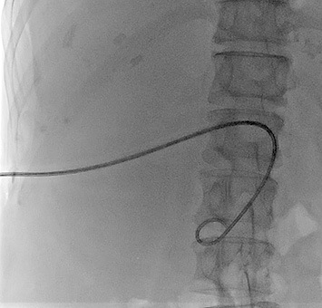
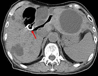
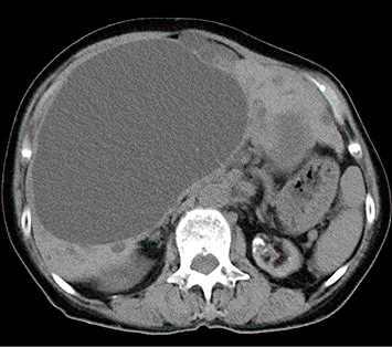
Herein, we have reported a rare case of intracystic hemorrhage due to rupture of a right hepatic artery pseudoaneurysm in a 76-year-old female patient who underwent drainage and 3% polidocanol sclerotherapy for a symptomatic giant hepatic cyst. One month after sclerotherapy, the patient presented to the emergency room with acute and severe abdominal pain. Non-contrast T1-weighted magnetic resonance imaging findings showed high hepatic cyst fluid signal intensity and abdominal arteriography findings revealed a right hepatic artery pseudoaneurysm surrounding the hepatic cystic wall. Therefore, the patient was diagnosed with intracystic hemorrhage due to a ruptured pseudoaneurysm. Embolization, using a detachable coil, was successful. Interventional radiologists should be aware of potential vascular injuries during drainage and sclerotherapy for giant hepatic cysts.


 分享
分享

 求助内容:
求助内容: 应助结果提醒方式:
 扫码关注我们
