Satoru Kawakita, Shaopei Li, Huu Tuan Nguyen, Surjendu Maity, Reihaneh Haghniaz, Jamal Bahari, Ning Yu, Kalpana Mandal, Praveen Bandaru, Lei Mou, Menekse Ermis, Enam Khalil, Safoora Khosravi, Arne Peirsman, Rohollah Nasiri, Annie Adachi, Aya Nakayama, Remy Bell, Yangzhi Zhu, Vadim Jucaud, Mehmet Remzi Dokmeci, Ali Khademhosseini
{"title":"将丝网印刷电极快速集成到热塑性芯片上的有机物设备中,用于实时监测跨内皮电阻。","authors":"Satoru Kawakita, Shaopei Li, Huu Tuan Nguyen, Surjendu Maity, Reihaneh Haghniaz, Jamal Bahari, Ning Yu, Kalpana Mandal, Praveen Bandaru, Lei Mou, Menekse Ermis, Enam Khalil, Safoora Khosravi, Arne Peirsman, Rohollah Nasiri, Annie Adachi, Aya Nakayama, Remy Bell, Yangzhi Zhu, Vadim Jucaud, Mehmet Remzi Dokmeci, Ali Khademhosseini","doi":"10.1007/s10544-023-00669-9","DOIUrl":null,"url":null,"abstract":"<div><p>Trans-endothelial electrical resistance (TEER) is one of the most widely used indicators to quantify the barrier integrity of endothelial layers. Over the last decade, the integration of TEER sensors into organ-on-a-chip (OOC) platforms has gained increasing interest for its efficient and effective measurement of TEER in OOCs. To date, microfabricated electrodes or direct insertion of wires has been used to integrate TEER sensors into OOCs, with each method having advantages and disadvantages. In this study, we developed a TEER-SPE chip consisting of carbon-based screen-printed electrodes (SPEs) embedded in a poly(methyl methacrylate) (PMMA)-based multi-layered microfluidic device with a porous poly(ethylene terephthalate) membrane in-between. As proof of concept, we demonstrated the successful cultures of hCMEC/D3 cells and the formation of confluent monolayers in the TEER-SPE chip and obtained TEER measurements for 4 days. Additionally, the TEER-SPE chip could detect changes in the barrier integrity due to shear stress or an inflammatory cytokine (i.e., tumor necrosis factor-α). The novel approach enables a low-cost and facile fabrication of carbon-based SPEs on PMMA substrates and the subsequent assembly of PMMA layers for rapid prototyping. Being cost-effective and cleanroom-free, our method lowers the existing logistical and technical barriers presenting itself as another step forward to the broader adoption of OOCs with TEER measurement capability.</p></div>","PeriodicalId":490,"journal":{"name":"Biomedical Microdevices","volume":"25 4","pages":""},"PeriodicalIF":3.3000,"publicationDate":"2023-09-23","publicationTypes":"Journal Article","fieldsOfStudy":null,"isOpenAccess":false,"openAccessPdf":"","citationCount":"0","resultStr":"{\"title\":\"Rapid integration of screen-printed electrodes into thermoplastic organ-on-a-chip devices for real-time monitoring of trans-endothelial electrical resistance\",\"authors\":\"Satoru Kawakita, Shaopei Li, Huu Tuan Nguyen, Surjendu Maity, Reihaneh Haghniaz, Jamal Bahari, Ning Yu, Kalpana Mandal, Praveen Bandaru, Lei Mou, Menekse Ermis, Enam Khalil, Safoora Khosravi, Arne Peirsman, Rohollah Nasiri, Annie Adachi, Aya Nakayama, Remy Bell, Yangzhi Zhu, Vadim Jucaud, Mehmet Remzi Dokmeci, Ali Khademhosseini\",\"doi\":\"10.1007/s10544-023-00669-9\",\"DOIUrl\":null,\"url\":null,\"abstract\":\"<div><p>Trans-endothelial electrical resistance (TEER) is one of the most widely used indicators to quantify the barrier integrity of endothelial layers. Over the last decade, the integration of TEER sensors into organ-on-a-chip (OOC) platforms has gained increasing interest for its efficient and effective measurement of TEER in OOCs. To date, microfabricated electrodes or direct insertion of wires has been used to integrate TEER sensors into OOCs, with each method having advantages and disadvantages. In this study, we developed a TEER-SPE chip consisting of carbon-based screen-printed electrodes (SPEs) embedded in a poly(methyl methacrylate) (PMMA)-based multi-layered microfluidic device with a porous poly(ethylene terephthalate) membrane in-between. As proof of concept, we demonstrated the successful cultures of hCMEC/D3 cells and the formation of confluent monolayers in the TEER-SPE chip and obtained TEER measurements for 4 days. Additionally, the TEER-SPE chip could detect changes in the barrier integrity due to shear stress or an inflammatory cytokine (i.e., tumor necrosis factor-α). The novel approach enables a low-cost and facile fabrication of carbon-based SPEs on PMMA substrates and the subsequent assembly of PMMA layers for rapid prototyping. Being cost-effective and cleanroom-free, our method lowers the existing logistical and technical barriers presenting itself as another step forward to the broader adoption of OOCs with TEER measurement capability.</p></div>\",\"PeriodicalId\":490,\"journal\":{\"name\":\"Biomedical Microdevices\",\"volume\":\"25 4\",\"pages\":\"\"},\"PeriodicalIF\":3.3000,\"publicationDate\":\"2023-09-23\",\"publicationTypes\":\"Journal Article\",\"fieldsOfStudy\":null,\"isOpenAccess\":false,\"openAccessPdf\":\"\",\"citationCount\":\"0\",\"resultStr\":null,\"platform\":\"Semanticscholar\",\"paperid\":null,\"PeriodicalName\":\"Biomedical Microdevices\",\"FirstCategoryId\":\"5\",\"ListUrlMain\":\"https://link.springer.com/article/10.1007/s10544-023-00669-9\",\"RegionNum\":4,\"RegionCategory\":\"医学\",\"ArticlePicture\":[],\"TitleCN\":null,\"AbstractTextCN\":null,\"PMCID\":null,\"EPubDate\":\"\",\"PubModel\":\"\",\"JCR\":\"Q3\",\"JCRName\":\"ENGINEERING, BIOMEDICAL\",\"Score\":null,\"Total\":0}","platform":"Semanticscholar","paperid":null,"PeriodicalName":"Biomedical Microdevices","FirstCategoryId":"5","ListUrlMain":"https://link.springer.com/article/10.1007/s10544-023-00669-9","RegionNum":4,"RegionCategory":"医学","ArticlePicture":[],"TitleCN":null,"AbstractTextCN":null,"PMCID":null,"EPubDate":"","PubModel":"","JCR":"Q3","JCRName":"ENGINEERING, BIOMEDICAL","Score":null,"Total":0}
Rapid integration of screen-printed electrodes into thermoplastic organ-on-a-chip devices for real-time monitoring of trans-endothelial electrical resistance
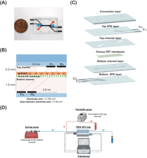
Trans-endothelial electrical resistance (TEER) is one of the most widely used indicators to quantify the barrier integrity of endothelial layers. Over the last decade, the integration of TEER sensors into organ-on-a-chip (OOC) platforms has gained increasing interest for its efficient and effective measurement of TEER in OOCs. To date, microfabricated electrodes or direct insertion of wires has been used to integrate TEER sensors into OOCs, with each method having advantages and disadvantages. In this study, we developed a TEER-SPE chip consisting of carbon-based screen-printed electrodes (SPEs) embedded in a poly(methyl methacrylate) (PMMA)-based multi-layered microfluidic device with a porous poly(ethylene terephthalate) membrane in-between. As proof of concept, we demonstrated the successful cultures of hCMEC/D3 cells and the formation of confluent monolayers in the TEER-SPE chip and obtained TEER measurements for 4 days. Additionally, the TEER-SPE chip could detect changes in the barrier integrity due to shear stress or an inflammatory cytokine (i.e., tumor necrosis factor-α). The novel approach enables a low-cost and facile fabrication of carbon-based SPEs on PMMA substrates and the subsequent assembly of PMMA layers for rapid prototyping. Being cost-effective and cleanroom-free, our method lowers the existing logistical and technical barriers presenting itself as another step forward to the broader adoption of OOCs with TEER measurement capability.
期刊介绍:
Biomedical Microdevices: BioMEMS and Biomedical Nanotechnology is an interdisciplinary periodical devoted to all aspects of research in the medical diagnostic and therapeutic applications of Micro-Electro-Mechanical Systems (BioMEMS) and nanotechnology for medicine and biology.
General subjects of interest include the design, characterization, testing, modeling and clinical validation of microfabricated systems, and their integration on-chip and in larger functional units. The specific interests of the Journal include systems for neural stimulation and recording, bioseparation technologies such as nanofilters and electrophoretic equipment, miniaturized analytic and DNA identification systems, biosensors, and micro/nanotechnologies for cell and tissue research, tissue engineering, cell transplantation, and the controlled release of drugs and biological molecules.
Contributions reporting on fundamental and applied investigations of the material science, biochemistry, and physics of biomedical microdevices and nanotechnology are encouraged. A non-exhaustive list of fields of interest includes: nanoparticle synthesis, characterization, and validation of therapeutic or imaging efficacy in animal models; biocompatibility; biochemical modification of microfabricated devices, with reference to non-specific protein adsorption, and the active immobilization and patterning of proteins on micro/nanofabricated surfaces; the dynamics of fluids in micro-and-nano-fabricated channels; the electromechanical and structural response of micro/nanofabricated systems; the interactions of microdevices with cells and tissues, including biocompatibility and biodegradation studies; variations in the characteristics of the systems as a function of the micro/nanofabrication parameters.


 分享
分享
 求助内容:
求助内容: 应助结果提醒方式:
 扫码关注我们
