Ali-Reza Ketabi, Stefan Hassfeld, Hans-Christoph Lauer, Andree Piwowarczyk
{"title":"上颌窦侧壁牙槽窦动脉管的全景摄影和锥束计算机断层扫描的对比诊断。","authors":"Ali-Reza Ketabi, Stefan Hassfeld, Hans-Christoph Lauer, Andree Piwowarczyk","doi":"10.1186/s40729-023-00497-9","DOIUrl":null,"url":null,"abstract":"<p><strong>Purpose: </strong>Sinus lift operations are a tried and tested means of providing adequate implant prosthetics to patients with compromised jawbones. Knowledge of the arterial supply of the maxillary sinus region is essential for surgical treatment in this area. The aim of the present comparative study was to determine whether alveolar antral artery (AAA) canal can be diagnosed both in corresponding panoramic radiography (PR) and cone-beam computed tomography (CBCT).</p><p><strong>Methods: </strong>A total of 335 patients with 635 sites and corresponding maxillary sinus in both PR and CBCT were selected and examined for AAA canal visibility.</p><p><strong>Results: </strong>The visibility of the AAA canal was significantly higher in CBCT than in PR. A total of 154 (46.0%) AAA canals could be identified in the maxillary sinus on the right. However, only four (1.2%) of these were also visible in PR. The detected values of the AAA canals in the maxillary sinus on the left in the PR and CBCT images were similar to those of the right. While 164 AAA canals (49%) were observed in CBCT images, only 1 (0.3%) was identifiable in PR.</p><p><strong>Conclusions: </strong>The results show that CBCT can be recommended for visualising the AAA canal when surgically planning sinus augmentation procedures.</p>","PeriodicalId":14076,"journal":{"name":"International Journal of Implant Dentistry","volume":"9 1","pages":"30"},"PeriodicalIF":3.1000,"publicationDate":"2023-09-19","publicationTypes":"Journal Article","fieldsOfStudy":null,"isOpenAccess":false,"openAccessPdf":"https://www.ncbi.nlm.nih.gov/pmc/articles/PMC10509091/pdf/","citationCount":"0","resultStr":"{\"title\":\"Comparative diagnosis of the alveolar antral artery canal in the lateral maxillary sinus wall in corresponding panoramic radiography and cone-beam computed tomography.\",\"authors\":\"Ali-Reza Ketabi, Stefan Hassfeld, Hans-Christoph Lauer, Andree Piwowarczyk\",\"doi\":\"10.1186/s40729-023-00497-9\",\"DOIUrl\":null,\"url\":null,\"abstract\":\"<p><strong>Purpose: </strong>Sinus lift operations are a tried and tested means of providing adequate implant prosthetics to patients with compromised jawbones. Knowledge of the arterial supply of the maxillary sinus region is essential for surgical treatment in this area. The aim of the present comparative study was to determine whether alveolar antral artery (AAA) canal can be diagnosed both in corresponding panoramic radiography (PR) and cone-beam computed tomography (CBCT).</p><p><strong>Methods: </strong>A total of 335 patients with 635 sites and corresponding maxillary sinus in both PR and CBCT were selected and examined for AAA canal visibility.</p><p><strong>Results: </strong>The visibility of the AAA canal was significantly higher in CBCT than in PR. A total of 154 (46.0%) AAA canals could be identified in the maxillary sinus on the right. However, only four (1.2%) of these were also visible in PR. The detected values of the AAA canals in the maxillary sinus on the left in the PR and CBCT images were similar to those of the right. While 164 AAA canals (49%) were observed in CBCT images, only 1 (0.3%) was identifiable in PR.</p><p><strong>Conclusions: </strong>The results show that CBCT can be recommended for visualising the AAA canal when surgically planning sinus augmentation procedures.</p>\",\"PeriodicalId\":14076,\"journal\":{\"name\":\"International Journal of Implant Dentistry\",\"volume\":\"9 1\",\"pages\":\"30\"},\"PeriodicalIF\":3.1000,\"publicationDate\":\"2023-09-19\",\"publicationTypes\":\"Journal Article\",\"fieldsOfStudy\":null,\"isOpenAccess\":false,\"openAccessPdf\":\"https://www.ncbi.nlm.nih.gov/pmc/articles/PMC10509091/pdf/\",\"citationCount\":\"0\",\"resultStr\":null,\"platform\":\"Semanticscholar\",\"paperid\":null,\"PeriodicalName\":\"International Journal of Implant Dentistry\",\"FirstCategoryId\":\"3\",\"ListUrlMain\":\"https://doi.org/10.1186/s40729-023-00497-9\",\"RegionNum\":3,\"RegionCategory\":\"医学\",\"ArticlePicture\":[],\"TitleCN\":null,\"AbstractTextCN\":null,\"PMCID\":null,\"EPubDate\":\"\",\"PubModel\":\"\",\"JCR\":\"Q1\",\"JCRName\":\"DENTISTRY, ORAL SURGERY & MEDICINE\",\"Score\":null,\"Total\":0}","platform":"Semanticscholar","paperid":null,"PeriodicalName":"International Journal of Implant Dentistry","FirstCategoryId":"3","ListUrlMain":"https://doi.org/10.1186/s40729-023-00497-9","RegionNum":3,"RegionCategory":"医学","ArticlePicture":[],"TitleCN":null,"AbstractTextCN":null,"PMCID":null,"EPubDate":"","PubModel":"","JCR":"Q1","JCRName":"DENTISTRY, ORAL SURGERY & MEDICINE","Score":null,"Total":0}
Comparative diagnosis of the alveolar antral artery canal in the lateral maxillary sinus wall in corresponding panoramic radiography and cone-beam computed tomography.
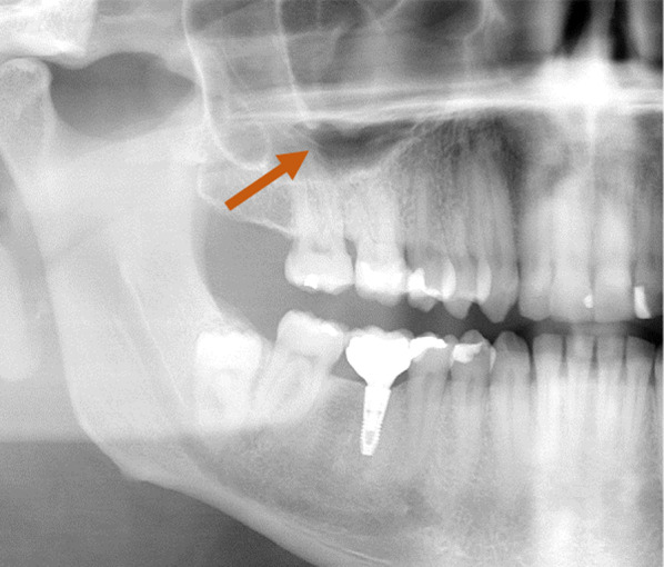
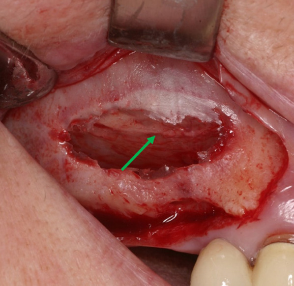
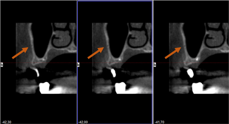
Purpose: Sinus lift operations are a tried and tested means of providing adequate implant prosthetics to patients with compromised jawbones. Knowledge of the arterial supply of the maxillary sinus region is essential for surgical treatment in this area. The aim of the present comparative study was to determine whether alveolar antral artery (AAA) canal can be diagnosed both in corresponding panoramic radiography (PR) and cone-beam computed tomography (CBCT).
Methods: A total of 335 patients with 635 sites and corresponding maxillary sinus in both PR and CBCT were selected and examined for AAA canal visibility.
Results: The visibility of the AAA canal was significantly higher in CBCT than in PR. A total of 154 (46.0%) AAA canals could be identified in the maxillary sinus on the right. However, only four (1.2%) of these were also visible in PR. The detected values of the AAA canals in the maxillary sinus on the left in the PR and CBCT images were similar to those of the right. While 164 AAA canals (49%) were observed in CBCT images, only 1 (0.3%) was identifiable in PR.
Conclusions: The results show that CBCT can be recommended for visualising the AAA canal when surgically planning sinus augmentation procedures.
期刊介绍:
The International Journal of Implant Dentistry is a peer-reviewed open access journal published under the SpringerOpen brand. The journal is dedicated to promoting the exchange and discussion of all research areas relevant to implant dentistry in the form of systematic literature or invited reviews, prospective and retrospective clinical studies, clinical case reports, basic laboratory and animal research, and articles on material research and engineering.


 分享
分享
 求助内容:
求助内容: 应助结果提醒方式:
 扫码关注我们
