Andrea Bianconi, Luca Francesco Rossi, Marta Bonada, Pietro Zeppa, Elsa Nico, Raffaele De Marco, Paola Lacroce, Fabio Cofano, Francesco Bruno, Giovanni Morana, Antonio Melcarne, Roberta Ruda, Luca Mainardi, Pietro Fiaschi, Diego Garbossa, Lia Morra
{"title":"基于深度学习的胶质母细胞瘤术后MRI分割算法:一种很有前途的肿瘤负担评估新工具。","authors":"Andrea Bianconi, Luca Francesco Rossi, Marta Bonada, Pietro Zeppa, Elsa Nico, Raffaele De Marco, Paola Lacroce, Fabio Cofano, Francesco Bruno, Giovanni Morana, Antonio Melcarne, Roberta Ruda, Luca Mainardi, Pietro Fiaschi, Diego Garbossa, Lia Morra","doi":"10.1186/s40708-023-00207-6","DOIUrl":null,"url":null,"abstract":"<p><strong>Objective: </strong>Clinical and surgical decisions for glioblastoma patients depend on a tumor imaging-based evaluation. Artificial Intelligence (AI) can be applied to magnetic resonance imaging (MRI) assessment to support clinical practice, surgery planning and prognostic predictions. In a real-world context, the current obstacles for AI are low-quality imaging and postoperative reliability. The aim of this study is to train an automatic algorithm for glioblastoma segmentation on a clinical MRI dataset and to obtain reliable results both pre- and post-operatively.</p><p><strong>Methods: </strong>The dataset used for this study comprises 237 (71 preoperative and 166 postoperative) MRIs from 71 patients affected by a histologically confirmed Grade IV Glioma. The implemented U-Net architecture was trained by transfer learning to perform the segmentation task on postoperative MRIs. The training was carried out first on BraTS2021 dataset for preoperative segmentation. Performance is evaluated using DICE score (DS) and Hausdorff 95% (H95).</p><p><strong>Results: </strong>In preoperative scenario, overall DS is 91.09 (± 0.60) and H95 is 8.35 (± 1.12), considering tumor core, enhancing tumor and whole tumor (ET and edema). In postoperative context, overall DS is 72.31 (± 2.88) and H95 is 23.43 (± 7.24), considering resection cavity (RC), gross tumor volume (GTV) and whole tumor (WT). Remarkably, the RC segmentation obtained a mean DS of 63.52 (± 8.90) in postoperative MRIs.</p><p><strong>Conclusions: </strong>The performances achieved by the algorithm are consistent with previous literature for both pre-operative and post-operative glioblastoma's MRI evaluation. Through the proposed algorithm, it is possible to reduce the impact of low-quality images and missing sequences.</p>","PeriodicalId":37465,"journal":{"name":"Brain Informatics","volume":"10 1","pages":"26"},"PeriodicalIF":4.5000,"publicationDate":"2023-10-06","publicationTypes":"Journal Article","fieldsOfStudy":null,"isOpenAccess":false,"openAccessPdf":"https://www.ncbi.nlm.nih.gov/pmc/articles/PMC10558414/pdf/","citationCount":"0","resultStr":"{\"title\":\"Deep learning-based algorithm for postoperative glioblastoma MRI segmentation: a promising new tool for tumor burden assessment.\",\"authors\":\"Andrea Bianconi, Luca Francesco Rossi, Marta Bonada, Pietro Zeppa, Elsa Nico, Raffaele De Marco, Paola Lacroce, Fabio Cofano, Francesco Bruno, Giovanni Morana, Antonio Melcarne, Roberta Ruda, Luca Mainardi, Pietro Fiaschi, Diego Garbossa, Lia Morra\",\"doi\":\"10.1186/s40708-023-00207-6\",\"DOIUrl\":null,\"url\":null,\"abstract\":\"<p><strong>Objective: </strong>Clinical and surgical decisions for glioblastoma patients depend on a tumor imaging-based evaluation. Artificial Intelligence (AI) can be applied to magnetic resonance imaging (MRI) assessment to support clinical practice, surgery planning and prognostic predictions. In a real-world context, the current obstacles for AI are low-quality imaging and postoperative reliability. The aim of this study is to train an automatic algorithm for glioblastoma segmentation on a clinical MRI dataset and to obtain reliable results both pre- and post-operatively.</p><p><strong>Methods: </strong>The dataset used for this study comprises 237 (71 preoperative and 166 postoperative) MRIs from 71 patients affected by a histologically confirmed Grade IV Glioma. The implemented U-Net architecture was trained by transfer learning to perform the segmentation task on postoperative MRIs. The training was carried out first on BraTS2021 dataset for preoperative segmentation. Performance is evaluated using DICE score (DS) and Hausdorff 95% (H95).</p><p><strong>Results: </strong>In preoperative scenario, overall DS is 91.09 (± 0.60) and H95 is 8.35 (± 1.12), considering tumor core, enhancing tumor and whole tumor (ET and edema). In postoperative context, overall DS is 72.31 (± 2.88) and H95 is 23.43 (± 7.24), considering resection cavity (RC), gross tumor volume (GTV) and whole tumor (WT). Remarkably, the RC segmentation obtained a mean DS of 63.52 (± 8.90) in postoperative MRIs.</p><p><strong>Conclusions: </strong>The performances achieved by the algorithm are consistent with previous literature for both pre-operative and post-operative glioblastoma's MRI evaluation. Through the proposed algorithm, it is possible to reduce the impact of low-quality images and missing sequences.</p>\",\"PeriodicalId\":37465,\"journal\":{\"name\":\"Brain Informatics\",\"volume\":\"10 1\",\"pages\":\"26\"},\"PeriodicalIF\":4.5000,\"publicationDate\":\"2023-10-06\",\"publicationTypes\":\"Journal Article\",\"fieldsOfStudy\":null,\"isOpenAccess\":false,\"openAccessPdf\":\"https://www.ncbi.nlm.nih.gov/pmc/articles/PMC10558414/pdf/\",\"citationCount\":\"0\",\"resultStr\":null,\"platform\":\"Semanticscholar\",\"paperid\":null,\"PeriodicalName\":\"Brain Informatics\",\"FirstCategoryId\":\"1085\",\"ListUrlMain\":\"https://doi.org/10.1186/s40708-023-00207-6\",\"RegionNum\":0,\"RegionCategory\":null,\"ArticlePicture\":[],\"TitleCN\":null,\"AbstractTextCN\":null,\"PMCID\":null,\"EPubDate\":\"\",\"PubModel\":\"\",\"JCR\":\"Q1\",\"JCRName\":\"Computer Science\",\"Score\":null,\"Total\":0}","platform":"Semanticscholar","paperid":null,"PeriodicalName":"Brain Informatics","FirstCategoryId":"1085","ListUrlMain":"https://doi.org/10.1186/s40708-023-00207-6","RegionNum":0,"RegionCategory":null,"ArticlePicture":[],"TitleCN":null,"AbstractTextCN":null,"PMCID":null,"EPubDate":"","PubModel":"","JCR":"Q1","JCRName":"Computer Science","Score":null,"Total":0}
Deep learning-based algorithm for postoperative glioblastoma MRI segmentation: a promising new tool for tumor burden assessment.
Objective: Clinical and surgical decisions for glioblastoma patients depend on a tumor imaging-based evaluation. Artificial Intelligence (AI) can be applied to magnetic resonance imaging (MRI) assessment to support clinical practice, surgery planning and prognostic predictions. In a real-world context, the current obstacles for AI are low-quality imaging and postoperative reliability. The aim of this study is to train an automatic algorithm for glioblastoma segmentation on a clinical MRI dataset and to obtain reliable results both pre- and post-operatively.
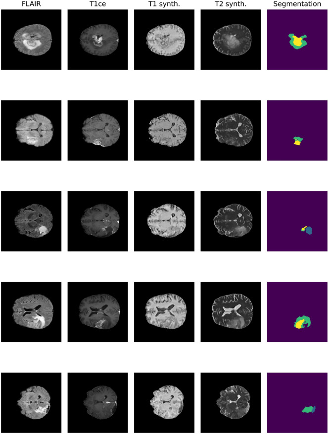
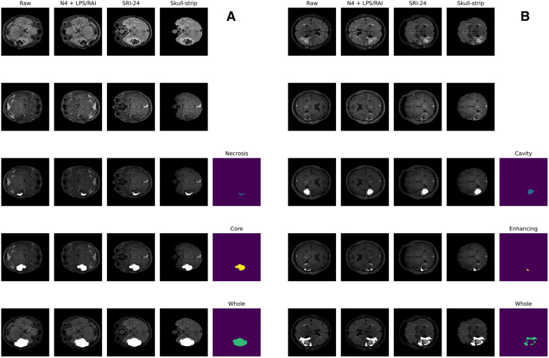
Methods: The dataset used for this study comprises 237 (71 preoperative and 166 postoperative) MRIs from 71 patients affected by a histologically confirmed Grade IV Glioma. The implemented U-Net architecture was trained by transfer learning to perform the segmentation task on postoperative MRIs. The training was carried out first on BraTS2021 dataset for preoperative segmentation. Performance is evaluated using DICE score (DS) and Hausdorff 95% (H95).
Results: In preoperative scenario, overall DS is 91.09 (± 0.60) and H95 is 8.35 (± 1.12), considering tumor core, enhancing tumor and whole tumor (ET and edema). In postoperative context, overall DS is 72.31 (± 2.88) and H95 is 23.43 (± 7.24), considering resection cavity (RC), gross tumor volume (GTV) and whole tumor (WT). Remarkably, the RC segmentation obtained a mean DS of 63.52 (± 8.90) in postoperative MRIs.
Conclusions: The performances achieved by the algorithm are consistent with previous literature for both pre-operative and post-operative glioblastoma's MRI evaluation. Through the proposed algorithm, it is possible to reduce the impact of low-quality images and missing sequences.
期刊介绍:
Brain Informatics is an international, peer-reviewed, interdisciplinary open-access journal published under the brand SpringerOpen, which provides a unique platform for researchers and practitioners to disseminate original research on computational and informatics technologies related to brain. This journal addresses the computational, cognitive, physiological, biological, physical, ecological and social perspectives of brain informatics. It also welcomes emerging information technologies and advanced neuro-imaging technologies, such as big data analytics and interactive knowledge discovery related to various large-scale brain studies and their applications. This journal will publish high-quality original research papers, brief reports and critical reviews in all theoretical, technological, clinical and interdisciplinary studies that make up the field of brain informatics and its applications in brain-machine intelligence, brain-inspired intelligent systems, mental health and brain disorders, etc. The scope of papers includes the following five tracks: Track 1: Cognitive and Computational Foundations of Brain Science Track 2: Human Information Processing Systems Track 3: Brain Big Data Analytics, Curation and Management Track 4: Informatics Paradigms for Brain and Mental Health Research Track 5: Brain-Machine Intelligence and Brain-Inspired Computing


 分享
分享
 求助内容:
求助内容: 应助结果提醒方式:
 扫码关注我们
