Laura Althea Cuschieri, Rebecca Schembri-Higgans, Nicholas Bezzina, Alexandra Betts, Arthur Rodriguez Gonzalez Cortes
{"title":"三维成像在软骨母细胞骨肉瘤早期诊断中的重要性。","authors":"Laura Althea Cuschieri, Rebecca Schembri-Higgans, Nicholas Bezzina, Alexandra Betts, Arthur Rodriguez Gonzalez Cortes","doi":"10.5624/isd.20220223","DOIUrl":null,"url":null,"abstract":"<p><p>The aim of this report is to present a case of chondroblastic osteosarcoma located in the right maxillary premolar region of a 17-year-old female patient. The initial clinical presentation and 2-dimensional (2D) radiographic methods proved inadequate for a definitive diagnosis. However, a cone-beam computed tomography scan revealed a hyperdense, heterogeneous lesion in the right maxillary premolar region, exhibiting a characteristic \"sun-ray\" appearance. To assess soft tissue involvement, a medical computed tomography scan was subsequently conducted. A positron emission tomography scan detected no metastasis or indications of secondary tumors. T1- and T2-weighted magnetic resonance imaging showed signal heterogeneity within the lesion, including areas of low signal intensity at the periphery. A histological examination conducted after an incisional biopsy confirmed the diagnosis of high-grade chondroblastic osteosarcoma. The patient was then referred to an oncology department for chemotherapy before surgery. In conclusion, these findings suggest that early diagnosis using 3-dimensional imaging can detect chondroblastic osteosarcoma in its early stages, such as before metastasis occurs, thereby improving the patient's prognosis.</p>","PeriodicalId":51714,"journal":{"name":"Imaging Science in Dentistry","volume":"53 3","pages":"247-256"},"PeriodicalIF":2.1000,"publicationDate":"2023-09-01","publicationTypes":"Journal Article","fieldsOfStudy":null,"isOpenAccess":false,"openAccessPdf":"https://ftp.ncbi.nlm.nih.gov/pub/pmc/oa_pdf/a0/19/isd-53-247.PMC10548150.pdf","citationCount":"0","resultStr":"{\"title\":\"Importance of 3-dimensional imaging in the early diagnosis of chondroblastic osteosarcoma.\",\"authors\":\"Laura Althea Cuschieri, Rebecca Schembri-Higgans, Nicholas Bezzina, Alexandra Betts, Arthur Rodriguez Gonzalez Cortes\",\"doi\":\"10.5624/isd.20220223\",\"DOIUrl\":null,\"url\":null,\"abstract\":\"<p><p>The aim of this report is to present a case of chondroblastic osteosarcoma located in the right maxillary premolar region of a 17-year-old female patient. The initial clinical presentation and 2-dimensional (2D) radiographic methods proved inadequate for a definitive diagnosis. However, a cone-beam computed tomography scan revealed a hyperdense, heterogeneous lesion in the right maxillary premolar region, exhibiting a characteristic \\\"sun-ray\\\" appearance. To assess soft tissue involvement, a medical computed tomography scan was subsequently conducted. A positron emission tomography scan detected no metastasis or indications of secondary tumors. T1- and T2-weighted magnetic resonance imaging showed signal heterogeneity within the lesion, including areas of low signal intensity at the periphery. A histological examination conducted after an incisional biopsy confirmed the diagnosis of high-grade chondroblastic osteosarcoma. The patient was then referred to an oncology department for chemotherapy before surgery. In conclusion, these findings suggest that early diagnosis using 3-dimensional imaging can detect chondroblastic osteosarcoma in its early stages, such as before metastasis occurs, thereby improving the patient's prognosis.</p>\",\"PeriodicalId\":51714,\"journal\":{\"name\":\"Imaging Science in Dentistry\",\"volume\":\"53 3\",\"pages\":\"247-256\"},\"PeriodicalIF\":2.1000,\"publicationDate\":\"2023-09-01\",\"publicationTypes\":\"Journal Article\",\"fieldsOfStudy\":null,\"isOpenAccess\":false,\"openAccessPdf\":\"https://ftp.ncbi.nlm.nih.gov/pub/pmc/oa_pdf/a0/19/isd-53-247.PMC10548150.pdf\",\"citationCount\":\"0\",\"resultStr\":null,\"platform\":\"Semanticscholar\",\"paperid\":null,\"PeriodicalName\":\"Imaging Science in Dentistry\",\"FirstCategoryId\":\"1085\",\"ListUrlMain\":\"https://doi.org/10.5624/isd.20220223\",\"RegionNum\":0,\"RegionCategory\":null,\"ArticlePicture\":[],\"TitleCN\":null,\"AbstractTextCN\":null,\"PMCID\":null,\"EPubDate\":\"2023/8/2 0:00:00\",\"PubModel\":\"Epub\",\"JCR\":\"Q3\",\"JCRName\":\"DENTISTRY, ORAL SURGERY & MEDICINE\",\"Score\":null,\"Total\":0}","platform":"Semanticscholar","paperid":null,"PeriodicalName":"Imaging Science in Dentistry","FirstCategoryId":"1085","ListUrlMain":"https://doi.org/10.5624/isd.20220223","RegionNum":0,"RegionCategory":null,"ArticlePicture":[],"TitleCN":null,"AbstractTextCN":null,"PMCID":null,"EPubDate":"2023/8/2 0:00:00","PubModel":"Epub","JCR":"Q3","JCRName":"DENTISTRY, ORAL SURGERY & MEDICINE","Score":null,"Total":0}
Importance of 3-dimensional imaging in the early diagnosis of chondroblastic osteosarcoma.
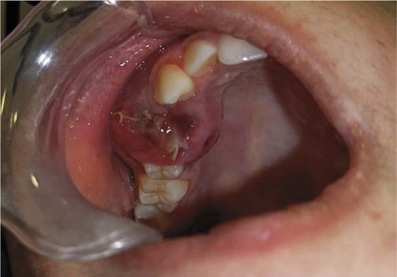
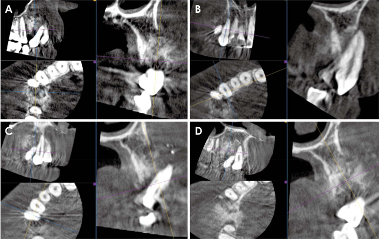
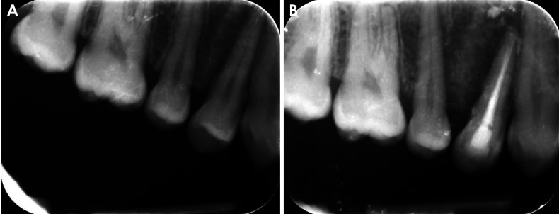
The aim of this report is to present a case of chondroblastic osteosarcoma located in the right maxillary premolar region of a 17-year-old female patient. The initial clinical presentation and 2-dimensional (2D) radiographic methods proved inadequate for a definitive diagnosis. However, a cone-beam computed tomography scan revealed a hyperdense, heterogeneous lesion in the right maxillary premolar region, exhibiting a characteristic "sun-ray" appearance. To assess soft tissue involvement, a medical computed tomography scan was subsequently conducted. A positron emission tomography scan detected no metastasis or indications of secondary tumors. T1- and T2-weighted magnetic resonance imaging showed signal heterogeneity within the lesion, including areas of low signal intensity at the periphery. A histological examination conducted after an incisional biopsy confirmed the diagnosis of high-grade chondroblastic osteosarcoma. The patient was then referred to an oncology department for chemotherapy before surgery. In conclusion, these findings suggest that early diagnosis using 3-dimensional imaging can detect chondroblastic osteosarcoma in its early stages, such as before metastasis occurs, thereby improving the patient's prognosis.


 分享
分享
 求助内容:
求助内容: 应助结果提醒方式:
 扫码关注我们
