{"title":"腮腺古代神经鞘瘤:一例报告并文献复习。","authors":"Young-Eun Kwon","doi":"10.5624/isd.20230504","DOIUrl":null,"url":null,"abstract":"<p><p>Schwannomas are uncommon neoplasms of neurologic origin that are rare in the salivary glands. A schwannoma that persists for a long time is referred to as an ancient schwannoma if it is accompanied by degenerative changes on histology. The case described herein involved a 37-year-old man with an ancient schwannoma that had persisted for 20 years in his right parotid gland. Clinically, the lesion presented with swelling and pain. Computed tomography revealed a well-defined, multilocular enhanced lesion. T2-weighted magnetic resonance images displayed multilocular hyperintensity, while T1-weighted images showed a high signal at the lobulated margin and a homogeneous low signal internally. The preoperative diagnosis, based on the lesion's location and imaging diagnosis, was Warthin's tumor. However, a biopsy conducted after surgical excision identified the lesion as a schwannoma with cystic degeneration. This report also presents a summary of the characteristics of rare cases of schwannoma in the major salivary gland based on this case and a literature review.</p>","PeriodicalId":51714,"journal":{"name":"Imaging Science in Dentistry","volume":"53 3","pages":"239-245"},"PeriodicalIF":2.1000,"publicationDate":"2023-09-01","publicationTypes":"Journal Article","fieldsOfStudy":null,"isOpenAccess":false,"openAccessPdf":"https://ftp.ncbi.nlm.nih.gov/pub/pmc/oa_pdf/b1/97/isd-53-239.PMC10548156.pdf","citationCount":"0","resultStr":"{\"title\":\"Ancient schwannoma in the parotid gland: A case report and review of the literature.\",\"authors\":\"Young-Eun Kwon\",\"doi\":\"10.5624/isd.20230504\",\"DOIUrl\":null,\"url\":null,\"abstract\":\"<p><p>Schwannomas are uncommon neoplasms of neurologic origin that are rare in the salivary glands. A schwannoma that persists for a long time is referred to as an ancient schwannoma if it is accompanied by degenerative changes on histology. The case described herein involved a 37-year-old man with an ancient schwannoma that had persisted for 20 years in his right parotid gland. Clinically, the lesion presented with swelling and pain. Computed tomography revealed a well-defined, multilocular enhanced lesion. T2-weighted magnetic resonance images displayed multilocular hyperintensity, while T1-weighted images showed a high signal at the lobulated margin and a homogeneous low signal internally. The preoperative diagnosis, based on the lesion's location and imaging diagnosis, was Warthin's tumor. However, a biopsy conducted after surgical excision identified the lesion as a schwannoma with cystic degeneration. This report also presents a summary of the characteristics of rare cases of schwannoma in the major salivary gland based on this case and a literature review.</p>\",\"PeriodicalId\":51714,\"journal\":{\"name\":\"Imaging Science in Dentistry\",\"volume\":\"53 3\",\"pages\":\"239-245\"},\"PeriodicalIF\":2.1000,\"publicationDate\":\"2023-09-01\",\"publicationTypes\":\"Journal Article\",\"fieldsOfStudy\":null,\"isOpenAccess\":false,\"openAccessPdf\":\"https://ftp.ncbi.nlm.nih.gov/pub/pmc/oa_pdf/b1/97/isd-53-239.PMC10548156.pdf\",\"citationCount\":\"0\",\"resultStr\":null,\"platform\":\"Semanticscholar\",\"paperid\":null,\"PeriodicalName\":\"Imaging Science in Dentistry\",\"FirstCategoryId\":\"1085\",\"ListUrlMain\":\"https://doi.org/10.5624/isd.20230504\",\"RegionNum\":0,\"RegionCategory\":null,\"ArticlePicture\":[],\"TitleCN\":null,\"AbstractTextCN\":null,\"PMCID\":null,\"EPubDate\":\"2023/9/4 0:00:00\",\"PubModel\":\"Epub\",\"JCR\":\"Q3\",\"JCRName\":\"DENTISTRY, ORAL SURGERY & MEDICINE\",\"Score\":null,\"Total\":0}","platform":"Semanticscholar","paperid":null,"PeriodicalName":"Imaging Science in Dentistry","FirstCategoryId":"1085","ListUrlMain":"https://doi.org/10.5624/isd.20230504","RegionNum":0,"RegionCategory":null,"ArticlePicture":[],"TitleCN":null,"AbstractTextCN":null,"PMCID":null,"EPubDate":"2023/9/4 0:00:00","PubModel":"Epub","JCR":"Q3","JCRName":"DENTISTRY, ORAL SURGERY & MEDICINE","Score":null,"Total":0}
Ancient schwannoma in the parotid gland: A case report and review of the literature.
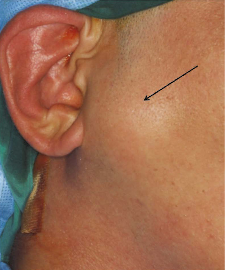
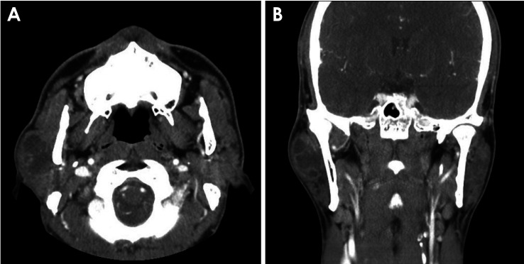
Schwannomas are uncommon neoplasms of neurologic origin that are rare in the salivary glands. A schwannoma that persists for a long time is referred to as an ancient schwannoma if it is accompanied by degenerative changes on histology. The case described herein involved a 37-year-old man with an ancient schwannoma that had persisted for 20 years in his right parotid gland. Clinically, the lesion presented with swelling and pain. Computed tomography revealed a well-defined, multilocular enhanced lesion. T2-weighted magnetic resonance images displayed multilocular hyperintensity, while T1-weighted images showed a high signal at the lobulated margin and a homogeneous low signal internally. The preoperative diagnosis, based on the lesion's location and imaging diagnosis, was Warthin's tumor. However, a biopsy conducted after surgical excision identified the lesion as a schwannoma with cystic degeneration. This report also presents a summary of the characteristics of rare cases of schwannoma in the major salivary gland based on this case and a literature review.


 分享
分享
 求助内容:
求助内容: 应助结果提醒方式:
 扫码关注我们
