Sami Lehtovirta, Victor Casula, Marianne Haapea, Simo Nortunen, Sannamari Lepojärvi, Harri Pakarinen, Miika T Nieminen, Eveliina Lammentausta, Jaakko Niinimäki
{"title":"使用T2放松时间评估稳定和不稳定的单侧weber型B/SER型踝关节骨折创伤后不久的踝关节关节软骨。","authors":"Sami Lehtovirta, Victor Casula, Marianne Haapea, Simo Nortunen, Sannamari Lepojärvi, Harri Pakarinen, Miika T Nieminen, Eveliina Lammentausta, Jaakko Niinimäki","doi":"10.1177/20584601231202033","DOIUrl":null,"url":null,"abstract":"<p><strong>Background: </strong>Early detection of post-traumatic cartilage damage in the ankle joint in magnetic resonance images can be difficult due to disturbances to structures usually appearing over time.</p><p><strong>Purpose: </strong>To study the articular cartilage of unilateral Weber type-B/SER-type ankle fractures shortly post-trauma using T2 relaxation time.</p><p><strong>Material and methods: </strong>Fifty one fractured ankles were gathered from consecutively screened patients, compiled initially for RCT studies, and treated at Oulu University Hospital and classified as stable (<i>n</i> = 28) and unstable fractures (<i>n</i> = 23) based on external-rotation stress test: medial clear space of ≥5 mm was interpreted as unstable. A control group of healthy young individuals (<i>n</i> = 19) was also gathered. All ankles were imaged on average 9 (range: 1 to 25) days after injury on a 3.0T MRI unit for T2 relaxation time assessment, and the cartilage was divided into sub-regions for comparison.</p><p><strong>Results: </strong>Control group displayed significantly higher T2 values in tibial cartilage compared to stable (six out of nine regions, <i>p</i>-values = .003-.043) and unstable (six out of nine regions, <i>p</i>-values = .001-.037) ankle fractures. No differences were detected in talar cartilage. Also, no differences were observed between stable and unstable fractures in tibial or talar cartilage.</p><p><strong>Conclusions: </strong>Lower T2 relaxation times of tibial cartilage in fractured ankles suggest intact extra cellular matrix (ECM) of the cartilage. Severity of the ankle fracture, measured by ankle stability, does not seem to increase ECM degradation immediately after trauma.</p>","PeriodicalId":72063,"journal":{"name":"Acta radiologica open","volume":"12 9","pages":"20584601231202033"},"PeriodicalIF":1.0000,"publicationDate":"2023-09-29","publicationTypes":"Journal Article","fieldsOfStudy":null,"isOpenAccess":false,"openAccessPdf":"https://www.ncbi.nlm.nih.gov/pmc/articles/PMC10540593/pdf/","citationCount":"0","resultStr":"{\"title\":\"Assessment of articular cartilage of ankle joint in stable and unstable unilateral weber type-B/SER-type ankle fractures shortly after trauma using T2 relaxation time.\",\"authors\":\"Sami Lehtovirta, Victor Casula, Marianne Haapea, Simo Nortunen, Sannamari Lepojärvi, Harri Pakarinen, Miika T Nieminen, Eveliina Lammentausta, Jaakko Niinimäki\",\"doi\":\"10.1177/20584601231202033\",\"DOIUrl\":null,\"url\":null,\"abstract\":\"<p><strong>Background: </strong>Early detection of post-traumatic cartilage damage in the ankle joint in magnetic resonance images can be difficult due to disturbances to structures usually appearing over time.</p><p><strong>Purpose: </strong>To study the articular cartilage of unilateral Weber type-B/SER-type ankle fractures shortly post-trauma using T2 relaxation time.</p><p><strong>Material and methods: </strong>Fifty one fractured ankles were gathered from consecutively screened patients, compiled initially for RCT studies, and treated at Oulu University Hospital and classified as stable (<i>n</i> = 28) and unstable fractures (<i>n</i> = 23) based on external-rotation stress test: medial clear space of ≥5 mm was interpreted as unstable. A control group of healthy young individuals (<i>n</i> = 19) was also gathered. All ankles were imaged on average 9 (range: 1 to 25) days after injury on a 3.0T MRI unit for T2 relaxation time assessment, and the cartilage was divided into sub-regions for comparison.</p><p><strong>Results: </strong>Control group displayed significantly higher T2 values in tibial cartilage compared to stable (six out of nine regions, <i>p</i>-values = .003-.043) and unstable (six out of nine regions, <i>p</i>-values = .001-.037) ankle fractures. No differences were detected in talar cartilage. Also, no differences were observed between stable and unstable fractures in tibial or talar cartilage.</p><p><strong>Conclusions: </strong>Lower T2 relaxation times of tibial cartilage in fractured ankles suggest intact extra cellular matrix (ECM) of the cartilage. Severity of the ankle fracture, measured by ankle stability, does not seem to increase ECM degradation immediately after trauma.</p>\",\"PeriodicalId\":72063,\"journal\":{\"name\":\"Acta radiologica open\",\"volume\":\"12 9\",\"pages\":\"20584601231202033\"},\"PeriodicalIF\":1.0000,\"publicationDate\":\"2023-09-29\",\"publicationTypes\":\"Journal Article\",\"fieldsOfStudy\":null,\"isOpenAccess\":false,\"openAccessPdf\":\"https://www.ncbi.nlm.nih.gov/pmc/articles/PMC10540593/pdf/\",\"citationCount\":\"0\",\"resultStr\":null,\"platform\":\"Semanticscholar\",\"paperid\":null,\"PeriodicalName\":\"Acta radiologica open\",\"FirstCategoryId\":\"1085\",\"ListUrlMain\":\"https://doi.org/10.1177/20584601231202033\",\"RegionNum\":0,\"RegionCategory\":null,\"ArticlePicture\":[],\"TitleCN\":null,\"AbstractTextCN\":null,\"PMCID\":null,\"EPubDate\":\"2023/9/1 0:00:00\",\"PubModel\":\"eCollection\",\"JCR\":\"Q4\",\"JCRName\":\"RADIOLOGY, NUCLEAR MEDICINE & MEDICAL IMAGING\",\"Score\":null,\"Total\":0}","platform":"Semanticscholar","paperid":null,"PeriodicalName":"Acta radiologica open","FirstCategoryId":"1085","ListUrlMain":"https://doi.org/10.1177/20584601231202033","RegionNum":0,"RegionCategory":null,"ArticlePicture":[],"TitleCN":null,"AbstractTextCN":null,"PMCID":null,"EPubDate":"2023/9/1 0:00:00","PubModel":"eCollection","JCR":"Q4","JCRName":"RADIOLOGY, NUCLEAR MEDICINE & MEDICAL IMAGING","Score":null,"Total":0}
Assessment of articular cartilage of ankle joint in stable and unstable unilateral weber type-B/SER-type ankle fractures shortly after trauma using T2 relaxation time.
Background: Early detection of post-traumatic cartilage damage in the ankle joint in magnetic resonance images can be difficult due to disturbances to structures usually appearing over time.
Purpose: To study the articular cartilage of unilateral Weber type-B/SER-type ankle fractures shortly post-trauma using T2 relaxation time.
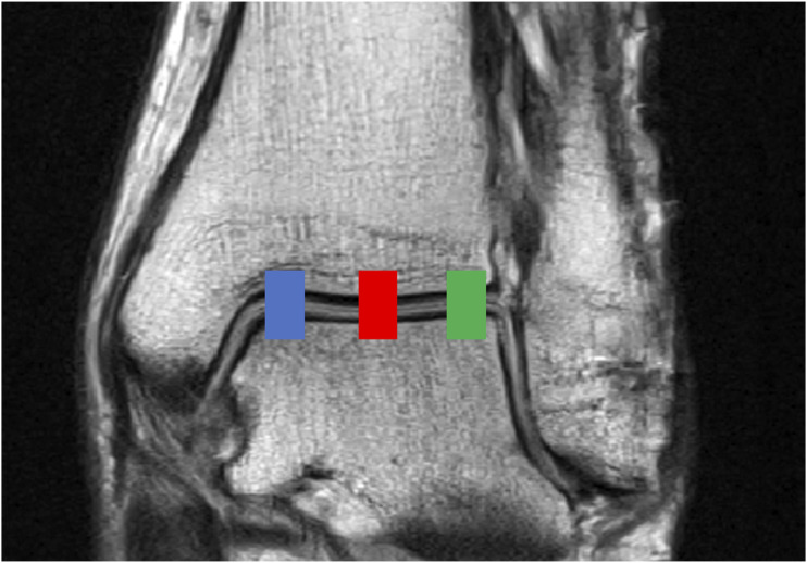
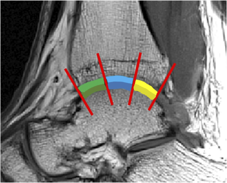
Material and methods: Fifty one fractured ankles were gathered from consecutively screened patients, compiled initially for RCT studies, and treated at Oulu University Hospital and classified as stable (n = 28) and unstable fractures (n = 23) based on external-rotation stress test: medial clear space of ≥5 mm was interpreted as unstable. A control group of healthy young individuals (n = 19) was also gathered. All ankles were imaged on average 9 (range: 1 to 25) days after injury on a 3.0T MRI unit for T2 relaxation time assessment, and the cartilage was divided into sub-regions for comparison.
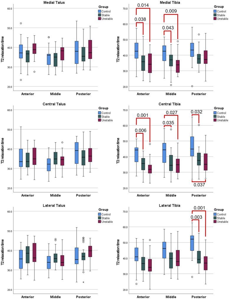
Results: Control group displayed significantly higher T2 values in tibial cartilage compared to stable (six out of nine regions, p-values = .003-.043) and unstable (six out of nine regions, p-values = .001-.037) ankle fractures. No differences were detected in talar cartilage. Also, no differences were observed between stable and unstable fractures in tibial or talar cartilage.
Conclusions: Lower T2 relaxation times of tibial cartilage in fractured ankles suggest intact extra cellular matrix (ECM) of the cartilage. Severity of the ankle fracture, measured by ankle stability, does not seem to increase ECM degradation immediately after trauma.


 分享
分享

 求助内容:
求助内容: 应助结果提醒方式:
 扫码关注我们
