Chen Lei, Jing Wang, Xin Li, Yuan-Yuan Mao, Jian-Qun Yan
{"title":"胰岛素抵抗高脂高糖饮食小鼠胰岛素受体的变化。","authors":"Chen Lei, Jing Wang, Xin Li, Yuan-Yuan Mao, Jian-Qun Yan","doi":"10.1080/21623945.2023.2264444","DOIUrl":null,"url":null,"abstract":"<p><p>This study aimed to observe the expression of insulin-signaling molecules in different organs of mice with insulin resistance (IR). Firstly, mice were fed a high-fat and high-sugar diet (HF group) to establish an IR model, and the controls (NF group) were fed with a normal diet. Next, the weight, fasting blood glucose (FBG), serum insulin and insulin tolerance were detected. Pathological changes of liver tissues were observed by H&E staining. The expressions of INSR, IRS-1 and IRS-2 in the liver, skeletal muscle and ovary were measured by qRT-PCR and western blotting. As a result, compared with the NF group, the HF group mice had increased weight, FBG, insulin and IR index after 6-week of feeding as well as a worse performance in the insulin tolerance test and H&E staining showed fatty liver-like changes after 12-week of feeding, exhibited lower expression of INSR, IRS-1 and IRS-2 in the liver of mice at 6 and 12 weeks. The expression of INSR and IRS-1 in skeletal muscle tissues exhibited the same trend, while those in ovary organs showed the opposite trend. These results suggested that the insulin signaling alters in the liver, skeletal muscle and ovary organs with the progress of IR.</p>","PeriodicalId":7226,"journal":{"name":"Adipocyte","volume":"12 1","pages":"2264444"},"PeriodicalIF":3.1000,"publicationDate":"2023-12-01","publicationTypes":"Journal Article","fieldsOfStudy":null,"isOpenAccess":false,"openAccessPdf":"https://www.ncbi.nlm.nih.gov/pmc/articles/PMC10578188/pdf/","citationCount":"1","resultStr":"{\"title\":\"Changes of insulin receptors in high fat and high glucose diet mice with insulin resistance.\",\"authors\":\"Chen Lei, Jing Wang, Xin Li, Yuan-Yuan Mao, Jian-Qun Yan\",\"doi\":\"10.1080/21623945.2023.2264444\",\"DOIUrl\":null,\"url\":null,\"abstract\":\"<p><p>This study aimed to observe the expression of insulin-signaling molecules in different organs of mice with insulin resistance (IR). Firstly, mice were fed a high-fat and high-sugar diet (HF group) to establish an IR model, and the controls (NF group) were fed with a normal diet. Next, the weight, fasting blood glucose (FBG), serum insulin and insulin tolerance were detected. Pathological changes of liver tissues were observed by H&E staining. The expressions of INSR, IRS-1 and IRS-2 in the liver, skeletal muscle and ovary were measured by qRT-PCR and western blotting. As a result, compared with the NF group, the HF group mice had increased weight, FBG, insulin and IR index after 6-week of feeding as well as a worse performance in the insulin tolerance test and H&E staining showed fatty liver-like changes after 12-week of feeding, exhibited lower expression of INSR, IRS-1 and IRS-2 in the liver of mice at 6 and 12 weeks. The expression of INSR and IRS-1 in skeletal muscle tissues exhibited the same trend, while those in ovary organs showed the opposite trend. These results suggested that the insulin signaling alters in the liver, skeletal muscle and ovary organs with the progress of IR.</p>\",\"PeriodicalId\":7226,\"journal\":{\"name\":\"Adipocyte\",\"volume\":\"12 1\",\"pages\":\"2264444\"},\"PeriodicalIF\":3.1000,\"publicationDate\":\"2023-12-01\",\"publicationTypes\":\"Journal Article\",\"fieldsOfStudy\":null,\"isOpenAccess\":false,\"openAccessPdf\":\"https://www.ncbi.nlm.nih.gov/pmc/articles/PMC10578188/pdf/\",\"citationCount\":\"1\",\"resultStr\":null,\"platform\":\"Semanticscholar\",\"paperid\":null,\"PeriodicalName\":\"Adipocyte\",\"FirstCategoryId\":\"99\",\"ListUrlMain\":\"https://doi.org/10.1080/21623945.2023.2264444\",\"RegionNum\":4,\"RegionCategory\":\"生物学\",\"ArticlePicture\":[],\"TitleCN\":null,\"AbstractTextCN\":null,\"PMCID\":null,\"EPubDate\":\"2023/10/13 0:00:00\",\"PubModel\":\"Epub\",\"JCR\":\"Q2\",\"JCRName\":\"ENDOCRINOLOGY & METABOLISM\",\"Score\":null,\"Total\":0}","platform":"Semanticscholar","paperid":null,"PeriodicalName":"Adipocyte","FirstCategoryId":"99","ListUrlMain":"https://doi.org/10.1080/21623945.2023.2264444","RegionNum":4,"RegionCategory":"生物学","ArticlePicture":[],"TitleCN":null,"AbstractTextCN":null,"PMCID":null,"EPubDate":"2023/10/13 0:00:00","PubModel":"Epub","JCR":"Q2","JCRName":"ENDOCRINOLOGY & METABOLISM","Score":null,"Total":0}
Changes of insulin receptors in high fat and high glucose diet mice with insulin resistance.
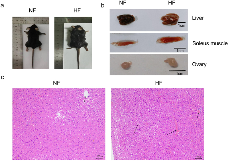
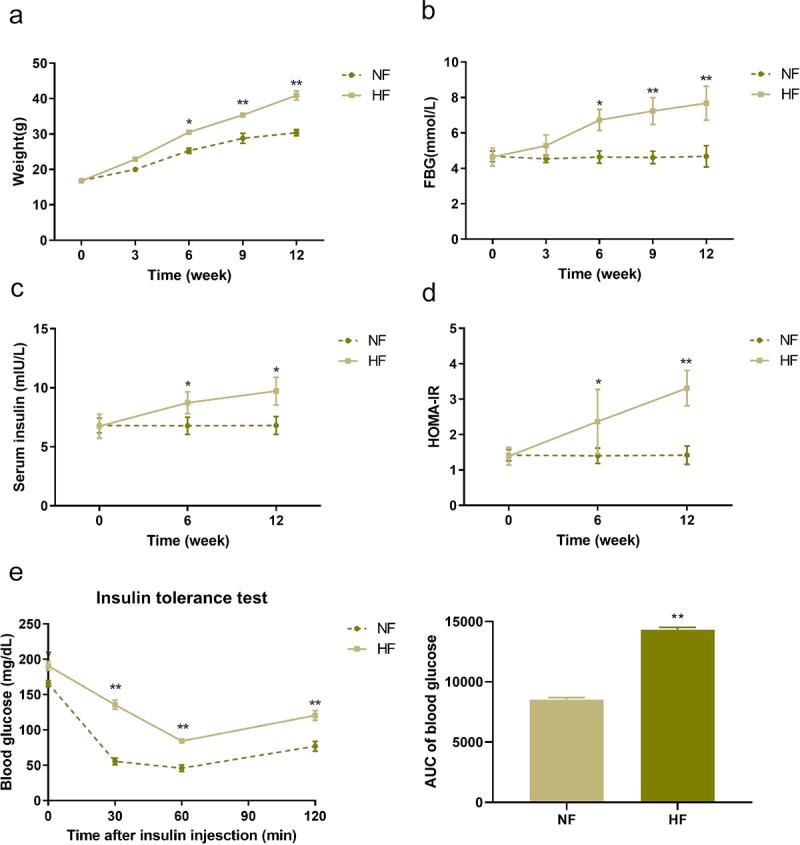
This study aimed to observe the expression of insulin-signaling molecules in different organs of mice with insulin resistance (IR). Firstly, mice were fed a high-fat and high-sugar diet (HF group) to establish an IR model, and the controls (NF group) were fed with a normal diet. Next, the weight, fasting blood glucose (FBG), serum insulin and insulin tolerance were detected. Pathological changes of liver tissues were observed by H&E staining. The expressions of INSR, IRS-1 and IRS-2 in the liver, skeletal muscle and ovary were measured by qRT-PCR and western blotting. As a result, compared with the NF group, the HF group mice had increased weight, FBG, insulin and IR index after 6-week of feeding as well as a worse performance in the insulin tolerance test and H&E staining showed fatty liver-like changes after 12-week of feeding, exhibited lower expression of INSR, IRS-1 and IRS-2 in the liver of mice at 6 and 12 weeks. The expression of INSR and IRS-1 in skeletal muscle tissues exhibited the same trend, while those in ovary organs showed the opposite trend. These results suggested that the insulin signaling alters in the liver, skeletal muscle and ovary organs with the progress of IR.
期刊介绍:
Adipocyte recognizes that the adipose tissue is the largest endocrine organ in the body, and explores the link between dysfunctional adipose tissue and the growing number of chronic diseases including diabetes, hypertension, cardiovascular disease and cancer. Historically, the primary function of the adipose tissue was limited to energy storage and thermoregulation. However, a plethora of research over the past 3 decades has recognized the dynamic role of the adipose tissue and its contribution to a variety of physiological processes including reproduction, angiogenesis, apoptosis, inflammation, blood pressure, coagulation, fibrinolysis, immunity and general metabolic homeostasis. The field of Adipose Tissue research has grown tremendously, and Adipocyte is the first international peer-reviewed journal of its kind providing a multi-disciplinary forum for research focusing exclusively on all aspects of adipose tissue physiology and pathophysiology. Adipocyte accepts high-profile submissions in basic, translational and clinical research.


 分享
分享
 求助内容:
求助内容: 应助结果提醒方式:
 扫码关注我们
