Rutika Sansaria , Krishanu Dey Das , Alwin Poulose
{"title":"使用机器学习模型量化高尔基体的扩散和分类。","authors":"Rutika Sansaria , Krishanu Dey Das , Alwin Poulose","doi":"10.1016/j.micron.2023.103547","DOIUrl":null,"url":null,"abstract":"<div><p>The Golgi body is a critical organelle in eukaryotic cells responsible for processing and modifying proteins and lipids. Under certain conditions, such as stress, disease, or ageing, the Golgi structure alters. Therefore, understanding the mechanisms that regulate Golgi dispersion has significant research contributions to identifying disease. However, there is a lack of tools to quantify the Golgi dispersion datasets. In this paper, we aim to automate the process of quantification of Golgi dispersion and use extracted features to classify dispersed Golgi images from undispersed Golgi images using machine learning models. First, we collected confocal microscopy images of transiently transfected HeLa cells expressing Galactose-1-phosphate uridylyltransferase (GALT)- green fluorescent protein (GFP) to quantify Golgi dispersal and classification. For the quantification, we introduced automated image processing and segmentation by applying mean and Gaussian filters. Then we used Otsu thresholding on preprocessed images and watershed segmentation to refine the segmentation of dispersed Golgi particles. In the case of classification, we extracted features from the Golgi dispersal images and classified them into empty vector (EV) versus CARP1 ring mutant (CARP1 RM) and empty vector (EV) versus CARP1 wildtype (CARP1 WT) classes. Our approach used machine-learning models, including logistic regression, decision tree, random forest, Naive Bayes, k-Nearest Neighbor (KNN), and gradient boosting for dispersed Golgi image classification. The experiment results show that our quantification technique on Golgi dispersal images reached 65% classification accuracy when the system uses a gradient boosting classifier for EV vs. CARP1 WT classification. Furthermore, our approach achieved 65% classification accuracy using a random forest classifier for EV vs. CARP1 RM classification.</p></div>","PeriodicalId":18501,"journal":{"name":"Micron","volume":"176 ","pages":"Article 103547"},"PeriodicalIF":2.2000,"publicationDate":"2024-01-01","publicationTypes":"Journal Article","fieldsOfStudy":null,"isOpenAccess":false,"openAccessPdf":"","citationCount":"0","resultStr":"{\"title\":\"Quantification of golgi dispersal and classification using machine learning models\",\"authors\":\"Rutika Sansaria , Krishanu Dey Das , Alwin Poulose\",\"doi\":\"10.1016/j.micron.2023.103547\",\"DOIUrl\":null,\"url\":null,\"abstract\":\"<div><p>The Golgi body is a critical organelle in eukaryotic cells responsible for processing and modifying proteins and lipids. Under certain conditions, such as stress, disease, or ageing, the Golgi structure alters. Therefore, understanding the mechanisms that regulate Golgi dispersion has significant research contributions to identifying disease. However, there is a lack of tools to quantify the Golgi dispersion datasets. In this paper, we aim to automate the process of quantification of Golgi dispersion and use extracted features to classify dispersed Golgi images from undispersed Golgi images using machine learning models. First, we collected confocal microscopy images of transiently transfected HeLa cells expressing Galactose-1-phosphate uridylyltransferase (GALT)- green fluorescent protein (GFP) to quantify Golgi dispersal and classification. For the quantification, we introduced automated image processing and segmentation by applying mean and Gaussian filters. Then we used Otsu thresholding on preprocessed images and watershed segmentation to refine the segmentation of dispersed Golgi particles. In the case of classification, we extracted features from the Golgi dispersal images and classified them into empty vector (EV) versus CARP1 ring mutant (CARP1 RM) and empty vector (EV) versus CARP1 wildtype (CARP1 WT) classes. Our approach used machine-learning models, including logistic regression, decision tree, random forest, Naive Bayes, k-Nearest Neighbor (KNN), and gradient boosting for dispersed Golgi image classification. The experiment results show that our quantification technique on Golgi dispersal images reached 65% classification accuracy when the system uses a gradient boosting classifier for EV vs. CARP1 WT classification. Furthermore, our approach achieved 65% classification accuracy using a random forest classifier for EV vs. CARP1 RM classification.</p></div>\",\"PeriodicalId\":18501,\"journal\":{\"name\":\"Micron\",\"volume\":\"176 \",\"pages\":\"Article 103547\"},\"PeriodicalIF\":2.2000,\"publicationDate\":\"2024-01-01\",\"publicationTypes\":\"Journal Article\",\"fieldsOfStudy\":null,\"isOpenAccess\":false,\"openAccessPdf\":\"\",\"citationCount\":\"0\",\"resultStr\":null,\"platform\":\"Semanticscholar\",\"paperid\":null,\"PeriodicalName\":\"Micron\",\"FirstCategoryId\":\"5\",\"ListUrlMain\":\"https://www.sciencedirect.com/science/article/pii/S0968432823001452\",\"RegionNum\":3,\"RegionCategory\":\"工程技术\",\"ArticlePicture\":[],\"TitleCN\":null,\"AbstractTextCN\":null,\"PMCID\":null,\"EPubDate\":\"2023/10/1 0:00:00\",\"PubModel\":\"Epub\",\"JCR\":\"Q1\",\"JCRName\":\"MICROSCOPY\",\"Score\":null,\"Total\":0}","platform":"Semanticscholar","paperid":null,"PeriodicalName":"Micron","FirstCategoryId":"5","ListUrlMain":"https://www.sciencedirect.com/science/article/pii/S0968432823001452","RegionNum":3,"RegionCategory":"工程技术","ArticlePicture":[],"TitleCN":null,"AbstractTextCN":null,"PMCID":null,"EPubDate":"2023/10/1 0:00:00","PubModel":"Epub","JCR":"Q1","JCRName":"MICROSCOPY","Score":null,"Total":0}
Quantification of golgi dispersal and classification using machine learning models
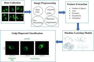
The Golgi body is a critical organelle in eukaryotic cells responsible for processing and modifying proteins and lipids. Under certain conditions, such as stress, disease, or ageing, the Golgi structure alters. Therefore, understanding the mechanisms that regulate Golgi dispersion has significant research contributions to identifying disease. However, there is a lack of tools to quantify the Golgi dispersion datasets. In this paper, we aim to automate the process of quantification of Golgi dispersion and use extracted features to classify dispersed Golgi images from undispersed Golgi images using machine learning models. First, we collected confocal microscopy images of transiently transfected HeLa cells expressing Galactose-1-phosphate uridylyltransferase (GALT)- green fluorescent protein (GFP) to quantify Golgi dispersal and classification. For the quantification, we introduced automated image processing and segmentation by applying mean and Gaussian filters. Then we used Otsu thresholding on preprocessed images and watershed segmentation to refine the segmentation of dispersed Golgi particles. In the case of classification, we extracted features from the Golgi dispersal images and classified them into empty vector (EV) versus CARP1 ring mutant (CARP1 RM) and empty vector (EV) versus CARP1 wildtype (CARP1 WT) classes. Our approach used machine-learning models, including logistic regression, decision tree, random forest, Naive Bayes, k-Nearest Neighbor (KNN), and gradient boosting for dispersed Golgi image classification. The experiment results show that our quantification technique on Golgi dispersal images reached 65% classification accuracy when the system uses a gradient boosting classifier for EV vs. CARP1 WT classification. Furthermore, our approach achieved 65% classification accuracy using a random forest classifier for EV vs. CARP1 RM classification.
期刊介绍:
Micron is an interdisciplinary forum for all work that involves new applications of microscopy or where advanced microscopy plays a central role. The journal will publish on the design, methods, application, practice or theory of microscopy and microanalysis, including reports on optical, electron-beam, X-ray microtomography, and scanning-probe systems. It also aims at the regular publication of review papers, short communications, as well as thematic issues on contemporary developments in microscopy and microanalysis. The journal embraces original research in which microscopy has contributed significantly to knowledge in biology, life science, nanoscience and nanotechnology, materials science and engineering.


 分享
分享
 求助内容:
求助内容: 应助结果提醒方式:
 扫码关注我们
