{"title":"径向结硅微柱阵列的深度依赖EBIC显微镜","authors":"Kaden M. Powell, Heayoung P. Yoon","doi":"10.1186/s42649-020-00037-4","DOIUrl":null,"url":null,"abstract":"<p>Recent advances in fabrication have enabled radial-junction architectures for cost-effective and high-performance optoelectronic devices. Unlike a planar PN junction, a radial-junction geometry maximizes the optical interaction in the three-dimensional (3D) structures, while effectively extracting the generated carriers via the conformal PN junction. In this paper, we report characterizations of radial PN junctions that consist of <i>p</i>-type Si micropillars created by deep reactive-ion etching (DRIE) and an <i>n</i>-type layer formed by phosphorus gas diffusion. We use electron-beam induced current (EBIC) microscopy to access the 3D junction profile from the sidewall of the pillars. Our EBIC images reveal uniform PN junctions conformally constructed on the 3D pillar array. Based on Monte-Carlo simulations and EBIC modeling, we estimate local carrier separation/collection efficiency that reflects the quality of the PN junction. We find the EBIC efficiency of the pillar array increases with the incident electron beam energy, consistent with the EBIC behaviors observed in a high-quality planar PN junction. The magnitude of the EBIC efficiency of our pillar array is about 70% at 10?kV, slightly lower than that of the planar device (≈ 81%). We suggest that this reduction could be attributed to the unpassivated pillar surface and the unintended recombination centers in the pillar cores introduced during the DRIE processes. Our results support that the depth-dependent EBIC approach is ideally suitable for evaluating PN junctions formed on micro/nanostructured semiconductors with various geometry.</p>","PeriodicalId":470,"journal":{"name":"Applied Microscopy","volume":"50 1","pages":""},"PeriodicalIF":0.0000,"publicationDate":"2020-09-03","publicationTypes":"Journal Article","fieldsOfStudy":null,"isOpenAccess":false,"openAccessPdf":"https://sci-hub-pdf.com/10.1186/s42649-020-00037-4","citationCount":"3","resultStr":"{\"title\":\"Depth-dependent EBIC microscopy of radial-junction Si micropillar arrays\",\"authors\":\"Kaden M. Powell, Heayoung P. Yoon\",\"doi\":\"10.1186/s42649-020-00037-4\",\"DOIUrl\":null,\"url\":null,\"abstract\":\"<p>Recent advances in fabrication have enabled radial-junction architectures for cost-effective and high-performance optoelectronic devices. Unlike a planar PN junction, a radial-junction geometry maximizes the optical interaction in the three-dimensional (3D) structures, while effectively extracting the generated carriers via the conformal PN junction. In this paper, we report characterizations of radial PN junctions that consist of <i>p</i>-type Si micropillars created by deep reactive-ion etching (DRIE) and an <i>n</i>-type layer formed by phosphorus gas diffusion. We use electron-beam induced current (EBIC) microscopy to access the 3D junction profile from the sidewall of the pillars. Our EBIC images reveal uniform PN junctions conformally constructed on the 3D pillar array. Based on Monte-Carlo simulations and EBIC modeling, we estimate local carrier separation/collection efficiency that reflects the quality of the PN junction. We find the EBIC efficiency of the pillar array increases with the incident electron beam energy, consistent with the EBIC behaviors observed in a high-quality planar PN junction. The magnitude of the EBIC efficiency of our pillar array is about 70% at 10?kV, slightly lower than that of the planar device (≈ 81%). We suggest that this reduction could be attributed to the unpassivated pillar surface and the unintended recombination centers in the pillar cores introduced during the DRIE processes. Our results support that the depth-dependent EBIC approach is ideally suitable for evaluating PN junctions formed on micro/nanostructured semiconductors with various geometry.</p>\",\"PeriodicalId\":470,\"journal\":{\"name\":\"Applied Microscopy\",\"volume\":\"50 1\",\"pages\":\"\"},\"PeriodicalIF\":0.0000,\"publicationDate\":\"2020-09-03\",\"publicationTypes\":\"Journal Article\",\"fieldsOfStudy\":null,\"isOpenAccess\":false,\"openAccessPdf\":\"https://sci-hub-pdf.com/10.1186/s42649-020-00037-4\",\"citationCount\":\"3\",\"resultStr\":null,\"platform\":\"Semanticscholar\",\"paperid\":null,\"PeriodicalName\":\"Applied Microscopy\",\"FirstCategoryId\":\"1085\",\"ListUrlMain\":\"https://link.springer.com/article/10.1186/s42649-020-00037-4\",\"RegionNum\":0,\"RegionCategory\":null,\"ArticlePicture\":[],\"TitleCN\":null,\"AbstractTextCN\":null,\"PMCID\":null,\"EPubDate\":\"\",\"PubModel\":\"\",\"JCR\":\"Q3\",\"JCRName\":\"Immunology and Microbiology\",\"Score\":null,\"Total\":0}","platform":"Semanticscholar","paperid":null,"PeriodicalName":"Applied Microscopy","FirstCategoryId":"1085","ListUrlMain":"https://link.springer.com/article/10.1186/s42649-020-00037-4","RegionNum":0,"RegionCategory":null,"ArticlePicture":[],"TitleCN":null,"AbstractTextCN":null,"PMCID":null,"EPubDate":"","PubModel":"","JCR":"Q3","JCRName":"Immunology and Microbiology","Score":null,"Total":0}
Depth-dependent EBIC microscopy of radial-junction Si micropillar arrays
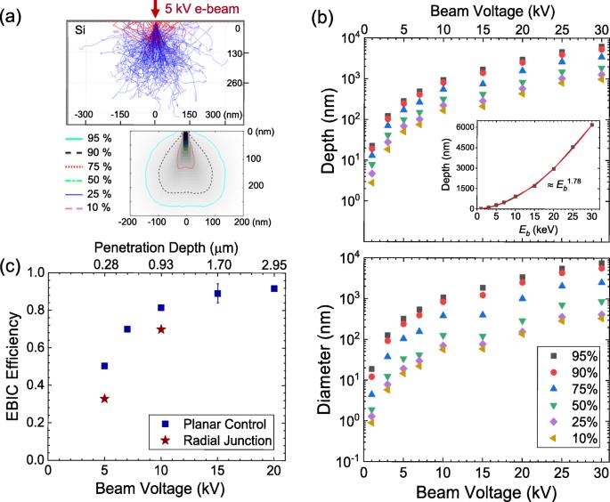
Recent advances in fabrication have enabled radial-junction architectures for cost-effective and high-performance optoelectronic devices. Unlike a planar PN junction, a radial-junction geometry maximizes the optical interaction in the three-dimensional (3D) structures, while effectively extracting the generated carriers via the conformal PN junction. In this paper, we report characterizations of radial PN junctions that consist of p-type Si micropillars created by deep reactive-ion etching (DRIE) and an n-type layer formed by phosphorus gas diffusion. We use electron-beam induced current (EBIC) microscopy to access the 3D junction profile from the sidewall of the pillars. Our EBIC images reveal uniform PN junctions conformally constructed on the 3D pillar array. Based on Monte-Carlo simulations and EBIC modeling, we estimate local carrier separation/collection efficiency that reflects the quality of the PN junction. We find the EBIC efficiency of the pillar array increases with the incident electron beam energy, consistent with the EBIC behaviors observed in a high-quality planar PN junction. The magnitude of the EBIC efficiency of our pillar array is about 70% at 10?kV, slightly lower than that of the planar device (≈ 81%). We suggest that this reduction could be attributed to the unpassivated pillar surface and the unintended recombination centers in the pillar cores introduced during the DRIE processes. Our results support that the depth-dependent EBIC approach is ideally suitable for evaluating PN junctions formed on micro/nanostructured semiconductors with various geometry.
Applied MicroscopyImmunology and Microbiology-Applied Microbiology and Biotechnology
CiteScore
3.40
自引率
0.00%
发文量
10
审稿时长
10 weeks
期刊介绍:
Applied Microscopy is a peer-reviewed journal sponsored by the Korean Society of Microscopy. The journal covers all the interdisciplinary fields of technological developments in new microscopy methods and instrumentation and their applications to biological or materials science for determining structure and chemistry. ISSN: 22875123, 22874445.


 分享
分享
 求助内容:
求助内容: 应助结果提醒方式:
 扫码关注我们
