Cosmin Adrian Teodoru, Maria-Emilia Cerghedean-Florea, Ciprian Tănăsescu, Horațiu Dura, Radu Fleacă, Mihnea Munteanu, Horia Stanca, Florina Georgeta Popescu, Mihai Dan Roman, Gheorghe Zsolt Nicula, Horea Vladi Matei, Mihaela Laura Vică
{"title":"白内障手术后眼部生物特征参数的评估","authors":"Cosmin Adrian Teodoru, Maria-Emilia Cerghedean-Florea, Ciprian Tănăsescu, Horațiu Dura, Radu Fleacă, Mihnea Munteanu, Horia Stanca, Florina Georgeta Popescu, Mihai Dan Roman, Gheorghe Zsolt Nicula, Horea Vladi Matei, Mihaela Laura Vică","doi":"10.3390/reports6010003","DOIUrl":null,"url":null,"abstract":"<p><strong>Background: </strong>The aim of this study was to highlight the structural changes in patients with cataract following surgery and the repercussions on the anterior pole.</p><p><strong>Methods: </strong>A total of 83 patients diagnosed with cataract who underwent uneventful phacoemulsification was included. Every patient was examined one week prior to and two weeks after the surgery. Pre- and postoperative assessment included examination of the anterior and posterior segment, keratometry, and optical biometry.</p><p><strong>Results: </strong>The pre- vs. postoperative axial length (AL) mean difference was 0.07 ± 0.18 mm (<i>p</i> < 0.001).The mean difference of the postoperative anterior chamber depth (ACD) vs. preoperative ACD values (1.11 ± 0.50 mm) was also statistically significant (<i>p</i> < 0.001). The linear regression function postoperative central corneal thickness (CCT) = 0.9004 × (preoperative CCT) + 0.0668, where it characterized a reduced positive correlation (R<sup>2</sup>) of 68.89% between the postoperative CCT and preoperative CCT. The mean pre-/post-operative differences in the K1 values were 0.08 ± 0.38 D, with a statistically significant difference between the two datasets (<i>p</i> = 0.0152). The mean pre/postoperative difference in the K2 values was 0.002 ± 0.58 D (<i>p</i> = 0.4854).</p><p><strong>Conclusions: </strong>ACD deepened significantly postoperatively. Regarding AL, there was a decrease after surgery, and a very good positive correlation between the post and preoperative values. The CCT values decreased with age. The 2.2-mm corneal incision during cataract surgery resulted in a relatively small postoperative residual astigmatism.</p>","PeriodicalId":74664,"journal":{"name":"Reports (MDPI)","volume":" ","pages":""},"PeriodicalIF":0.8000,"publicationDate":"2023-01-18","publicationTypes":"Journal Article","fieldsOfStudy":null,"isOpenAccess":false,"openAccessPdf":"https://www.ncbi.nlm.nih.gov/pmc/articles/PMC12225319/pdf/","citationCount":"0","resultStr":"{\"title\":\"Evaluation of Ocular Biometric Parameters Following Cataract Surgery.\",\"authors\":\"Cosmin Adrian Teodoru, Maria-Emilia Cerghedean-Florea, Ciprian Tănăsescu, Horațiu Dura, Radu Fleacă, Mihnea Munteanu, Horia Stanca, Florina Georgeta Popescu, Mihai Dan Roman, Gheorghe Zsolt Nicula, Horea Vladi Matei, Mihaela Laura Vică\",\"doi\":\"10.3390/reports6010003\",\"DOIUrl\":null,\"url\":null,\"abstract\":\"<p><strong>Background: </strong>The aim of this study was to highlight the structural changes in patients with cataract following surgery and the repercussions on the anterior pole.</p><p><strong>Methods: </strong>A total of 83 patients diagnosed with cataract who underwent uneventful phacoemulsification was included. Every patient was examined one week prior to and two weeks after the surgery. Pre- and postoperative assessment included examination of the anterior and posterior segment, keratometry, and optical biometry.</p><p><strong>Results: </strong>The pre- vs. postoperative axial length (AL) mean difference was 0.07 ± 0.18 mm (<i>p</i> < 0.001).The mean difference of the postoperative anterior chamber depth (ACD) vs. preoperative ACD values (1.11 ± 0.50 mm) was also statistically significant (<i>p</i> < 0.001). The linear regression function postoperative central corneal thickness (CCT) = 0.9004 × (preoperative CCT) + 0.0668, where it characterized a reduced positive correlation (R<sup>2</sup>) of 68.89% between the postoperative CCT and preoperative CCT. The mean pre-/post-operative differences in the K1 values were 0.08 ± 0.38 D, with a statistically significant difference between the two datasets (<i>p</i> = 0.0152). The mean pre/postoperative difference in the K2 values was 0.002 ± 0.58 D (<i>p</i> = 0.4854).</p><p><strong>Conclusions: </strong>ACD deepened significantly postoperatively. Regarding AL, there was a decrease after surgery, and a very good positive correlation between the post and preoperative values. The CCT values decreased with age. The 2.2-mm corneal incision during cataract surgery resulted in a relatively small postoperative residual astigmatism.</p>\",\"PeriodicalId\":74664,\"journal\":{\"name\":\"Reports (MDPI)\",\"volume\":\" \",\"pages\":\"\"},\"PeriodicalIF\":0.8000,\"publicationDate\":\"2023-01-18\",\"publicationTypes\":\"Journal Article\",\"fieldsOfStudy\":null,\"isOpenAccess\":false,\"openAccessPdf\":\"https://www.ncbi.nlm.nih.gov/pmc/articles/PMC12225319/pdf/\",\"citationCount\":\"0\",\"resultStr\":null,\"platform\":\"Semanticscholar\",\"paperid\":null,\"PeriodicalName\":\"Reports (MDPI)\",\"FirstCategoryId\":\"1085\",\"ListUrlMain\":\"https://doi.org/10.3390/reports6010003\",\"RegionNum\":0,\"RegionCategory\":null,\"ArticlePicture\":[],\"TitleCN\":null,\"AbstractTextCN\":null,\"PMCID\":null,\"EPubDate\":\"\",\"PubModel\":\"\",\"JCR\":\"Q3\",\"JCRName\":\"MEDICINE, GENERAL & INTERNAL\",\"Score\":null,\"Total\":0}","platform":"Semanticscholar","paperid":null,"PeriodicalName":"Reports (MDPI)","FirstCategoryId":"1085","ListUrlMain":"https://doi.org/10.3390/reports6010003","RegionNum":0,"RegionCategory":null,"ArticlePicture":[],"TitleCN":null,"AbstractTextCN":null,"PMCID":null,"EPubDate":"","PubModel":"","JCR":"Q3","JCRName":"MEDICINE, GENERAL & INTERNAL","Score":null,"Total":0}
Evaluation of Ocular Biometric Parameters Following Cataract Surgery.
Background: The aim of this study was to highlight the structural changes in patients with cataract following surgery and the repercussions on the anterior pole.
Methods: A total of 83 patients diagnosed with cataract who underwent uneventful phacoemulsification was included. Every patient was examined one week prior to and two weeks after the surgery. Pre- and postoperative assessment included examination of the anterior and posterior segment, keratometry, and optical biometry.
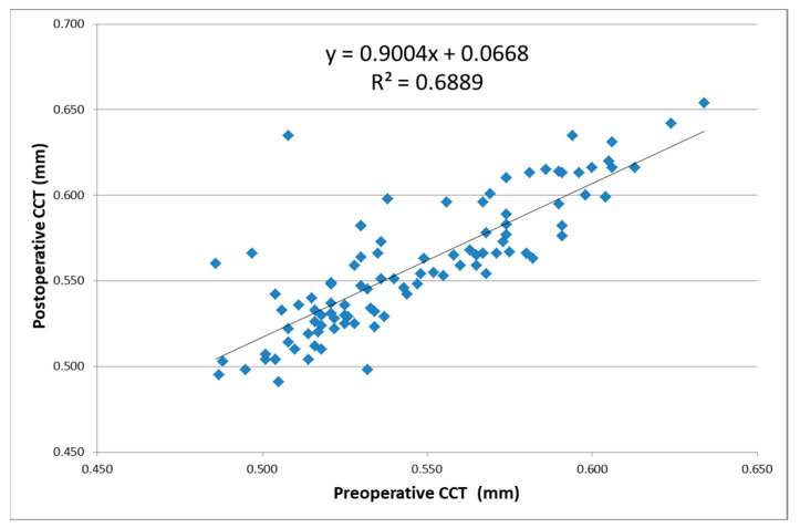
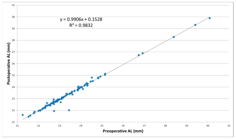
Results: The pre- vs. postoperative axial length (AL) mean difference was 0.07 ± 0.18 mm (p < 0.001).The mean difference of the postoperative anterior chamber depth (ACD) vs. preoperative ACD values (1.11 ± 0.50 mm) was also statistically significant (p < 0.001). The linear regression function postoperative central corneal thickness (CCT) = 0.9004 × (preoperative CCT) + 0.0668, where it characterized a reduced positive correlation (R2) of 68.89% between the postoperative CCT and preoperative CCT. The mean pre-/post-operative differences in the K1 values were 0.08 ± 0.38 D, with a statistically significant difference between the two datasets (p = 0.0152). The mean pre/postoperative difference in the K2 values was 0.002 ± 0.58 D (p = 0.4854).
Conclusions: ACD deepened significantly postoperatively. Regarding AL, there was a decrease after surgery, and a very good positive correlation between the post and preoperative values. The CCT values decreased with age. The 2.2-mm corneal incision during cataract surgery resulted in a relatively small postoperative residual astigmatism.


 分享
分享

 求助内容:
求助内容: 应助结果提醒方式:
 扫码关注我们
