{"title":"圆网蜘蛛心肌细胞的精细结构","authors":"Yan Sun, Hyo-Jeong Kim, Myung-Jin Moon","doi":"10.1186/s42649-020-00030-x","DOIUrl":null,"url":null,"abstract":"<p>The fine structural characteristics of cardiac muscle cells and its myofibril organization in the orb web spider <i>N. clavata</i> were examined by transmission electron microscopy. Although myofibril striations are not remarkable as those of skeletal muscles, muscle fibers contain multiple myofibrils, abundant mitochondria, extensive sarcoplasmic reticulum and transverse tubules (T-tubules). Myofibrils are divided into distinct sarcomeres defined by Z-lines with average length of 2.0?μm, but the distinction between the A-band and the I-bands is not clear due to uniform striations over the length of the sarcomeres. Dyadic junction which consisted of a single T-tubule paired with a terminal cisterna of the sarcoplasmic reticulum is found mainly at the A-I level of sarcomere. Each cell is arranged to form multiple connections with neighboring cells through the intercalated discs. These specialized junctions include three types of intercellular junctions: gap junctions, fascia adherens and desmosomes for heart function. Our transmission electron microscopy (TEM) observations clearly show that spider’s cardiac muscle contraction is controlled by neurogenic rather than myogenic mechanism since each cardiac muscle fiber is innervated by a branch of motor neuron through neuromuscular junctions.</p>","PeriodicalId":470,"journal":{"name":"Applied Microscopy","volume":"50 1","pages":""},"PeriodicalIF":0.0000,"publicationDate":"2020-05-14","publicationTypes":"Journal Article","fieldsOfStudy":null,"isOpenAccess":false,"openAccessPdf":"https://sci-hub-pdf.com/10.1186/s42649-020-00030-x","citationCount":"2","resultStr":"{\"title\":\"Fine structure of the cardiac muscle cells in the orb-web spider Nephila clavata\",\"authors\":\"Yan Sun, Hyo-Jeong Kim, Myung-Jin Moon\",\"doi\":\"10.1186/s42649-020-00030-x\",\"DOIUrl\":null,\"url\":null,\"abstract\":\"<p>The fine structural characteristics of cardiac muscle cells and its myofibril organization in the orb web spider <i>N. clavata</i> were examined by transmission electron microscopy. Although myofibril striations are not remarkable as those of skeletal muscles, muscle fibers contain multiple myofibrils, abundant mitochondria, extensive sarcoplasmic reticulum and transverse tubules (T-tubules). Myofibrils are divided into distinct sarcomeres defined by Z-lines with average length of 2.0?μm, but the distinction between the A-band and the I-bands is not clear due to uniform striations over the length of the sarcomeres. Dyadic junction which consisted of a single T-tubule paired with a terminal cisterna of the sarcoplasmic reticulum is found mainly at the A-I level of sarcomere. Each cell is arranged to form multiple connections with neighboring cells through the intercalated discs. These specialized junctions include three types of intercellular junctions: gap junctions, fascia adherens and desmosomes for heart function. Our transmission electron microscopy (TEM) observations clearly show that spider’s cardiac muscle contraction is controlled by neurogenic rather than myogenic mechanism since each cardiac muscle fiber is innervated by a branch of motor neuron through neuromuscular junctions.</p>\",\"PeriodicalId\":470,\"journal\":{\"name\":\"Applied Microscopy\",\"volume\":\"50 1\",\"pages\":\"\"},\"PeriodicalIF\":0.0000,\"publicationDate\":\"2020-05-14\",\"publicationTypes\":\"Journal Article\",\"fieldsOfStudy\":null,\"isOpenAccess\":false,\"openAccessPdf\":\"https://sci-hub-pdf.com/10.1186/s42649-020-00030-x\",\"citationCount\":\"2\",\"resultStr\":null,\"platform\":\"Semanticscholar\",\"paperid\":null,\"PeriodicalName\":\"Applied Microscopy\",\"FirstCategoryId\":\"1085\",\"ListUrlMain\":\"https://link.springer.com/article/10.1186/s42649-020-00030-x\",\"RegionNum\":0,\"RegionCategory\":null,\"ArticlePicture\":[],\"TitleCN\":null,\"AbstractTextCN\":null,\"PMCID\":null,\"EPubDate\":\"\",\"PubModel\":\"\",\"JCR\":\"Q3\",\"JCRName\":\"Immunology and Microbiology\",\"Score\":null,\"Total\":0}","platform":"Semanticscholar","paperid":null,"PeriodicalName":"Applied Microscopy","FirstCategoryId":"1085","ListUrlMain":"https://link.springer.com/article/10.1186/s42649-020-00030-x","RegionNum":0,"RegionCategory":null,"ArticlePicture":[],"TitleCN":null,"AbstractTextCN":null,"PMCID":null,"EPubDate":"","PubModel":"","JCR":"Q3","JCRName":"Immunology and Microbiology","Score":null,"Total":0}
Fine structure of the cardiac muscle cells in the orb-web spider Nephila clavata
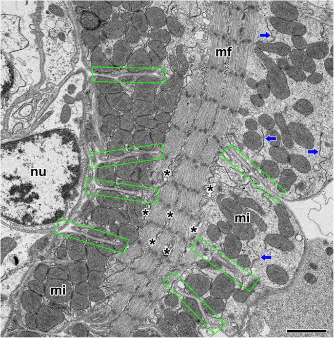
The fine structural characteristics of cardiac muscle cells and its myofibril organization in the orb web spider N. clavata were examined by transmission electron microscopy. Although myofibril striations are not remarkable as those of skeletal muscles, muscle fibers contain multiple myofibrils, abundant mitochondria, extensive sarcoplasmic reticulum and transverse tubules (T-tubules). Myofibrils are divided into distinct sarcomeres defined by Z-lines with average length of 2.0?μm, but the distinction between the A-band and the I-bands is not clear due to uniform striations over the length of the sarcomeres. Dyadic junction which consisted of a single T-tubule paired with a terminal cisterna of the sarcoplasmic reticulum is found mainly at the A-I level of sarcomere. Each cell is arranged to form multiple connections with neighboring cells through the intercalated discs. These specialized junctions include three types of intercellular junctions: gap junctions, fascia adherens and desmosomes for heart function. Our transmission electron microscopy (TEM) observations clearly show that spider’s cardiac muscle contraction is controlled by neurogenic rather than myogenic mechanism since each cardiac muscle fiber is innervated by a branch of motor neuron through neuromuscular junctions.
Applied MicroscopyImmunology and Microbiology-Applied Microbiology and Biotechnology
CiteScore
3.40
自引率
0.00%
发文量
10
审稿时长
10 weeks
期刊介绍:
Applied Microscopy is a peer-reviewed journal sponsored by the Korean Society of Microscopy. The journal covers all the interdisciplinary fields of technological developments in new microscopy methods and instrumentation and their applications to biological or materials science for determining structure and chemistry. ISSN: 22875123, 22874445.


 分享
分享
 求助内容:
求助内容: 应助结果提醒方式:
 扫码关注我们
