Raktim Bhattacharya, Sulagna Saha, Olga Kostina, Lyudmila Muravnik, Adinpunya Mitra
{"title":"用低成本的化学干燥法代替临界点干燥法,可以获得与之相当的扫描电镜图像质量","authors":"Raktim Bhattacharya, Sulagna Saha, Olga Kostina, Lyudmila Muravnik, Adinpunya Mitra","doi":"10.1186/s42649-020-00035-6","DOIUrl":null,"url":null,"abstract":"<p>Sample preparation including dehydration and drying of samples is the most intricate part of scanning electron microscopy. Most current sample preparation protocols use critical-point drying with liquid carbon dioxide. Very few studies have reported samples that were dried using chemical reagents. In this study, we used hexamethyldisilazane, a chemical drying reagent, to prepare plant samples. As glandular trichomes are among the most fragile and sensitive surface structures found on plants, we used <i>Millingtonia hortensis</i> leaf samples as our study materials because they contain abundant glandular trichomes. The results obtained using this new method are identical to those produced via critical-point drying.</p>","PeriodicalId":470,"journal":{"name":"Applied Microscopy","volume":"50 1","pages":""},"PeriodicalIF":0.0000,"publicationDate":"2020-07-17","publicationTypes":"Journal Article","fieldsOfStudy":null,"isOpenAccess":false,"openAccessPdf":"https://www.ncbi.nlm.nih.gov/pmc/articles/PMC7818294/pdf/","citationCount":"18","resultStr":"{\"title\":\"Replacing critical point drying with a low-cost chemical drying provides comparable surface image quality of glandular trichomes from leaves of Millingtonia hortensis L. f. in scanning electron micrograph\",\"authors\":\"Raktim Bhattacharya, Sulagna Saha, Olga Kostina, Lyudmila Muravnik, Adinpunya Mitra\",\"doi\":\"10.1186/s42649-020-00035-6\",\"DOIUrl\":null,\"url\":null,\"abstract\":\"<p>Sample preparation including dehydration and drying of samples is the most intricate part of scanning electron microscopy. Most current sample preparation protocols use critical-point drying with liquid carbon dioxide. Very few studies have reported samples that were dried using chemical reagents. In this study, we used hexamethyldisilazane, a chemical drying reagent, to prepare plant samples. As glandular trichomes are among the most fragile and sensitive surface structures found on plants, we used <i>Millingtonia hortensis</i> leaf samples as our study materials because they contain abundant glandular trichomes. The results obtained using this new method are identical to those produced via critical-point drying.</p>\",\"PeriodicalId\":470,\"journal\":{\"name\":\"Applied Microscopy\",\"volume\":\"50 1\",\"pages\":\"\"},\"PeriodicalIF\":0.0000,\"publicationDate\":\"2020-07-17\",\"publicationTypes\":\"Journal Article\",\"fieldsOfStudy\":null,\"isOpenAccess\":false,\"openAccessPdf\":\"https://www.ncbi.nlm.nih.gov/pmc/articles/PMC7818294/pdf/\",\"citationCount\":\"18\",\"resultStr\":null,\"platform\":\"Semanticscholar\",\"paperid\":null,\"PeriodicalName\":\"Applied Microscopy\",\"FirstCategoryId\":\"1085\",\"ListUrlMain\":\"https://link.springer.com/article/10.1186/s42649-020-00035-6\",\"RegionNum\":0,\"RegionCategory\":null,\"ArticlePicture\":[],\"TitleCN\":null,\"AbstractTextCN\":null,\"PMCID\":null,\"EPubDate\":\"\",\"PubModel\":\"\",\"JCR\":\"Q3\",\"JCRName\":\"Immunology and Microbiology\",\"Score\":null,\"Total\":0}","platform":"Semanticscholar","paperid":null,"PeriodicalName":"Applied Microscopy","FirstCategoryId":"1085","ListUrlMain":"https://link.springer.com/article/10.1186/s42649-020-00035-6","RegionNum":0,"RegionCategory":null,"ArticlePicture":[],"TitleCN":null,"AbstractTextCN":null,"PMCID":null,"EPubDate":"","PubModel":"","JCR":"Q3","JCRName":"Immunology and Microbiology","Score":null,"Total":0}
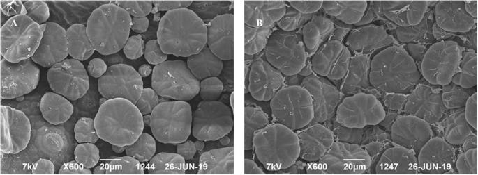
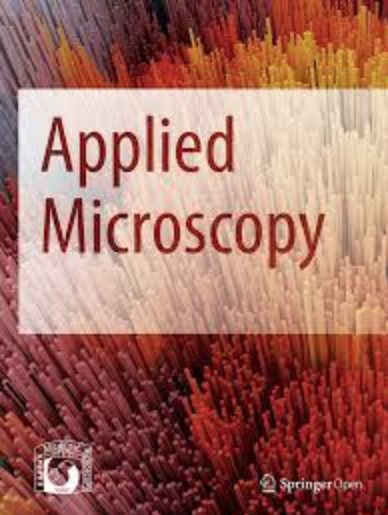
Replacing critical point drying with a low-cost chemical drying provides comparable surface image quality of glandular trichomes from leaves of Millingtonia hortensis L. f. in scanning electron micrograph
Sample preparation including dehydration and drying of samples is the most intricate part of scanning electron microscopy. Most current sample preparation protocols use critical-point drying with liquid carbon dioxide. Very few studies have reported samples that were dried using chemical reagents. In this study, we used hexamethyldisilazane, a chemical drying reagent, to prepare plant samples. As glandular trichomes are among the most fragile and sensitive surface structures found on plants, we used Millingtonia hortensis leaf samples as our study materials because they contain abundant glandular trichomes. The results obtained using this new method are identical to those produced via critical-point drying.
Applied MicroscopyImmunology and Microbiology-Applied Microbiology and Biotechnology
CiteScore
3.40
自引率
0.00%
发文量
10
审稿时长
10 weeks
期刊介绍:
Applied Microscopy is a peer-reviewed journal sponsored by the Korean Society of Microscopy. The journal covers all the interdisciplinary fields of technological developments in new microscopy methods and instrumentation and their applications to biological or materials science for determining structure and chemistry. ISSN: 22875123, 22874445.


 分享
分享
 求助内容:
求助内容: 应助结果提醒方式:
 扫码关注我们
