{"title":"荧光探针测定突触前功能的方法","authors":"Yeseul Jang, Sung Rae Kim, Sung Hoon Lee","doi":"10.1186/s42649-021-00051-0","DOIUrl":null,"url":null,"abstract":"<p>Synaptic vesicles, which are endogenous to neurotransmitters, are involved in exocytosis by active potentials and release neurotransmitters. Synaptic vesicles used in neurotransmitter release are reused via endocytosis to maintain a pool of synaptic vesicles. Synaptic vesicles show different types of exo- and endocytosis depending on animal species, type of nerve cell, and electrical activity. To accurately understand the dynamics of synaptic vesicles, direct observation of synaptic vesicles is required; however, it was difficult to observe synaptic vesicles of size 40–50?nm in living neurons. The exo-and endocytosis of synaptic vesicles was confirmed by labeling the vesicles with a fluorescent agent and measuring the changes in fluorescence intensity. To date, various methods of labeling synaptic vesicles have been proposed, and each method has its own characteristics, strength, and drawbacks. In this study, we introduce methods that can measure presynaptic activity and describe the characteristics of each technique.</p>","PeriodicalId":470,"journal":{"name":"Applied Microscopy","volume":"51 1","pages":""},"PeriodicalIF":0.0000,"publicationDate":"2021-03-17","publicationTypes":"Journal Article","fieldsOfStudy":null,"isOpenAccess":false,"openAccessPdf":"https://sci-hub-pdf.com/10.1186/s42649-021-00051-0","citationCount":"2","resultStr":"{\"title\":\"Methods of measuring presynaptic function with fluorescence probes\",\"authors\":\"Yeseul Jang, Sung Rae Kim, Sung Hoon Lee\",\"doi\":\"10.1186/s42649-021-00051-0\",\"DOIUrl\":null,\"url\":null,\"abstract\":\"<p>Synaptic vesicles, which are endogenous to neurotransmitters, are involved in exocytosis by active potentials and release neurotransmitters. Synaptic vesicles used in neurotransmitter release are reused via endocytosis to maintain a pool of synaptic vesicles. Synaptic vesicles show different types of exo- and endocytosis depending on animal species, type of nerve cell, and electrical activity. To accurately understand the dynamics of synaptic vesicles, direct observation of synaptic vesicles is required; however, it was difficult to observe synaptic vesicles of size 40–50?nm in living neurons. The exo-and endocytosis of synaptic vesicles was confirmed by labeling the vesicles with a fluorescent agent and measuring the changes in fluorescence intensity. To date, various methods of labeling synaptic vesicles have been proposed, and each method has its own characteristics, strength, and drawbacks. In this study, we introduce methods that can measure presynaptic activity and describe the characteristics of each technique.</p>\",\"PeriodicalId\":470,\"journal\":{\"name\":\"Applied Microscopy\",\"volume\":\"51 1\",\"pages\":\"\"},\"PeriodicalIF\":0.0000,\"publicationDate\":\"2021-03-17\",\"publicationTypes\":\"Journal Article\",\"fieldsOfStudy\":null,\"isOpenAccess\":false,\"openAccessPdf\":\"https://sci-hub-pdf.com/10.1186/s42649-021-00051-0\",\"citationCount\":\"2\",\"resultStr\":null,\"platform\":\"Semanticscholar\",\"paperid\":null,\"PeriodicalName\":\"Applied Microscopy\",\"FirstCategoryId\":\"1085\",\"ListUrlMain\":\"https://link.springer.com/article/10.1186/s42649-021-00051-0\",\"RegionNum\":0,\"RegionCategory\":null,\"ArticlePicture\":[],\"TitleCN\":null,\"AbstractTextCN\":null,\"PMCID\":null,\"EPubDate\":\"\",\"PubModel\":\"\",\"JCR\":\"Q3\",\"JCRName\":\"Immunology and Microbiology\",\"Score\":null,\"Total\":0}","platform":"Semanticscholar","paperid":null,"PeriodicalName":"Applied Microscopy","FirstCategoryId":"1085","ListUrlMain":"https://link.springer.com/article/10.1186/s42649-021-00051-0","RegionNum":0,"RegionCategory":null,"ArticlePicture":[],"TitleCN":null,"AbstractTextCN":null,"PMCID":null,"EPubDate":"","PubModel":"","JCR":"Q3","JCRName":"Immunology and Microbiology","Score":null,"Total":0}
Methods of measuring presynaptic function with fluorescence probes
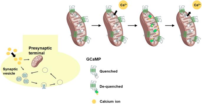
Synaptic vesicles, which are endogenous to neurotransmitters, are involved in exocytosis by active potentials and release neurotransmitters. Synaptic vesicles used in neurotransmitter release are reused via endocytosis to maintain a pool of synaptic vesicles. Synaptic vesicles show different types of exo- and endocytosis depending on animal species, type of nerve cell, and electrical activity. To accurately understand the dynamics of synaptic vesicles, direct observation of synaptic vesicles is required; however, it was difficult to observe synaptic vesicles of size 40–50?nm in living neurons. The exo-and endocytosis of synaptic vesicles was confirmed by labeling the vesicles with a fluorescent agent and measuring the changes in fluorescence intensity. To date, various methods of labeling synaptic vesicles have been proposed, and each method has its own characteristics, strength, and drawbacks. In this study, we introduce methods that can measure presynaptic activity and describe the characteristics of each technique.
Applied MicroscopyImmunology and Microbiology-Applied Microbiology and Biotechnology
CiteScore
3.40
自引率
0.00%
发文量
10
审稿时长
10 weeks
期刊介绍:
Applied Microscopy is a peer-reviewed journal sponsored by the Korean Society of Microscopy. The journal covers all the interdisciplinary fields of technological developments in new microscopy methods and instrumentation and their applications to biological or materials science for determining structure and chemistry. ISSN: 22875123, 22874445.


 分享
分享
 求助内容:
求助内容: 应助结果提醒方式:
 扫码关注我们
