Killivalavan Asaithambi , Iman Biswas , Kaza Suguna
{"title":"原生动物DNA损伤诱导蛋白1 (Ddi1)的结构和功能研究","authors":"Killivalavan Asaithambi , Iman Biswas , Kaza Suguna","doi":"10.1016/j.crstbi.2022.05.003","DOIUrl":null,"url":null,"abstract":"<div><p>Ddi1 is a multidomain protein that belongs to the ubiquitin receptor family of proteins. The Ddi1 proteins contain a highly conserved retroviral protease (RVP)-like domain along with other domains. The severity of opportunistic infections, caused by parasitic protozoa in AIDS patients, was found to decline when HIV protease inhibitors were used in antiretroviral therapy. Parasite growth was shown to be suppressed by a few of the inhibitors targeting Ddi1 present in these parasites. In this study, the binding of HIV protease inhibitors to the RVP domain of Ddi1 from <em>Toxoplasma gondii</em> and <em>Cryptosporidium hominis</em>; and the binding of ubiquitin to the ubiquitin-associated domain of Ddi1 from these two parasites were established using Biolayer Interferometry. The crystal structures of the RVP domains of Ddi1 from <em>T. gondii</em> and <em>C. hominis</em> were determined; they form homodimers similar to those observed in HIV protease and the reported structures of the same domain from <em>Saccharomyces cerevisiae</em>, <em>Leishmania major</em> and humans. The native form of the domain showed an open dimeric structure and a normal mode analysis revealed that it can take up a closed conformation resulting from relative movements of the subunits. Based on the crystal structure of the RVP domain of Ddi1 from <em>L. major</em>, a seven residue peptide inhibitor was designed and it was shown to bind to the RVP domain of Ddi1 from <em>L. major</em> by Biolayer Interferometry. This peptide was modified using computational methods and was shown to have a better affinity than the initial peptide.</p></div>","PeriodicalId":10870,"journal":{"name":"Current Research in Structural Biology","volume":"4 ","pages":"Pages 175-191"},"PeriodicalIF":2.7000,"publicationDate":"2022-01-01","publicationTypes":"Journal Article","fieldsOfStudy":null,"isOpenAccess":false,"openAccessPdf":"https://www.sciencedirect.com/science/article/pii/S2665928X22000150/pdfft?md5=71c62a69e55ec00d3ea56e5e57a30aac&pid=1-s2.0-S2665928X22000150-main.pdf","citationCount":"0","resultStr":"{\"title\":\"Structural and functional insights into the DNA damage-inducible protein 1 (Ddi1) from protozoa\",\"authors\":\"Killivalavan Asaithambi , Iman Biswas , Kaza Suguna\",\"doi\":\"10.1016/j.crstbi.2022.05.003\",\"DOIUrl\":null,\"url\":null,\"abstract\":\"<div><p>Ddi1 is a multidomain protein that belongs to the ubiquitin receptor family of proteins. The Ddi1 proteins contain a highly conserved retroviral protease (RVP)-like domain along with other domains. The severity of opportunistic infections, caused by parasitic protozoa in AIDS patients, was found to decline when HIV protease inhibitors were used in antiretroviral therapy. Parasite growth was shown to be suppressed by a few of the inhibitors targeting Ddi1 present in these parasites. In this study, the binding of HIV protease inhibitors to the RVP domain of Ddi1 from <em>Toxoplasma gondii</em> and <em>Cryptosporidium hominis</em>; and the binding of ubiquitin to the ubiquitin-associated domain of Ddi1 from these two parasites were established using Biolayer Interferometry. The crystal structures of the RVP domains of Ddi1 from <em>T. gondii</em> and <em>C. hominis</em> were determined; they form homodimers similar to those observed in HIV protease and the reported structures of the same domain from <em>Saccharomyces cerevisiae</em>, <em>Leishmania major</em> and humans. The native form of the domain showed an open dimeric structure and a normal mode analysis revealed that it can take up a closed conformation resulting from relative movements of the subunits. Based on the crystal structure of the RVP domain of Ddi1 from <em>L. major</em>, a seven residue peptide inhibitor was designed and it was shown to bind to the RVP domain of Ddi1 from <em>L. major</em> by Biolayer Interferometry. This peptide was modified using computational methods and was shown to have a better affinity than the initial peptide.</p></div>\",\"PeriodicalId\":10870,\"journal\":{\"name\":\"Current Research in Structural Biology\",\"volume\":\"4 \",\"pages\":\"Pages 175-191\"},\"PeriodicalIF\":2.7000,\"publicationDate\":\"2022-01-01\",\"publicationTypes\":\"Journal Article\",\"fieldsOfStudy\":null,\"isOpenAccess\":false,\"openAccessPdf\":\"https://www.sciencedirect.com/science/article/pii/S2665928X22000150/pdfft?md5=71c62a69e55ec00d3ea56e5e57a30aac&pid=1-s2.0-S2665928X22000150-main.pdf\",\"citationCount\":\"0\",\"resultStr\":null,\"platform\":\"Semanticscholar\",\"paperid\":null,\"PeriodicalName\":\"Current Research in Structural Biology\",\"FirstCategoryId\":\"1085\",\"ListUrlMain\":\"https://www.sciencedirect.com/science/article/pii/S2665928X22000150\",\"RegionNum\":0,\"RegionCategory\":null,\"ArticlePicture\":[],\"TitleCN\":null,\"AbstractTextCN\":null,\"PMCID\":null,\"EPubDate\":\"2022/5/26 0:00:00\",\"PubModel\":\"Epub\",\"JCR\":\"Q3\",\"JCRName\":\"BIOCHEMISTRY & MOLECULAR BIOLOGY\",\"Score\":null,\"Total\":0}","platform":"Semanticscholar","paperid":null,"PeriodicalName":"Current Research in Structural Biology","FirstCategoryId":"1085","ListUrlMain":"https://www.sciencedirect.com/science/article/pii/S2665928X22000150","RegionNum":0,"RegionCategory":null,"ArticlePicture":[],"TitleCN":null,"AbstractTextCN":null,"PMCID":null,"EPubDate":"2022/5/26 0:00:00","PubModel":"Epub","JCR":"Q3","JCRName":"BIOCHEMISTRY & MOLECULAR BIOLOGY","Score":null,"Total":0}
Structural and functional insights into the DNA damage-inducible protein 1 (Ddi1) from protozoa
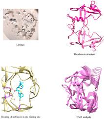
Ddi1 is a multidomain protein that belongs to the ubiquitin receptor family of proteins. The Ddi1 proteins contain a highly conserved retroviral protease (RVP)-like domain along with other domains. The severity of opportunistic infections, caused by parasitic protozoa in AIDS patients, was found to decline when HIV protease inhibitors were used in antiretroviral therapy. Parasite growth was shown to be suppressed by a few of the inhibitors targeting Ddi1 present in these parasites. In this study, the binding of HIV protease inhibitors to the RVP domain of Ddi1 from Toxoplasma gondii and Cryptosporidium hominis; and the binding of ubiquitin to the ubiquitin-associated domain of Ddi1 from these two parasites were established using Biolayer Interferometry. The crystal structures of the RVP domains of Ddi1 from T. gondii and C. hominis were determined; they form homodimers similar to those observed in HIV protease and the reported structures of the same domain from Saccharomyces cerevisiae, Leishmania major and humans. The native form of the domain showed an open dimeric structure and a normal mode analysis revealed that it can take up a closed conformation resulting from relative movements of the subunits. Based on the crystal structure of the RVP domain of Ddi1 from L. major, a seven residue peptide inhibitor was designed and it was shown to bind to the RVP domain of Ddi1 from L. major by Biolayer Interferometry. This peptide was modified using computational methods and was shown to have a better affinity than the initial peptide.


 分享
分享
 求助内容:
求助内容: 应助结果提醒方式:
 扫码关注我们
