{"title":"春季黑海西北陆架和深水区浮游植物的垂直分布","authors":"Vladimir Mukhanov , Evgeniy Sakhon , Natalia Rodionova , An-Yi Tsai","doi":"10.1016/j.jmarsys.2022.103779","DOIUrl":null,"url":null,"abstract":"<div><p><span>Structure and functions of picophytoplankton and their links to the water column density stratification and nutrient vertical profiles were studied in the Black Sea open waters along a transect from the Western Gyre to the NW shelf during spring homothermy in April of 2017. Abundances of picocyanobacteria of the genus </span><span><em>Synechococcus</em></span> (0.85 ± 0.96 (SD) × 10<sup>3</sup> cells ml<sup>−1</sup>) and eukaryotic picoalgae (5.74 ± 5.99 × 10<sup>3</sup> cells ml<sup>−1</sup>) and their intracellular pigment (chl <em>a</em>, phycoerythrin) contents were quantified using flow cytometry. The distribution of these variables in the upper 100-m layer was non-uniform. Along the whole transect, <em>Synechococcus</em><span> and picoeukaryote abundance maxima (up to 4 × 10</span><sup>3</sup> and 3 × 10<sup>4</sup> cells ml<sup>−1</sup>, respectively) were observed at the water temperature of 8–8.5°С in the depth range between 30 and 40 m. Picoeukaryotes dominated the total community. The share of <em>Synecococcus</em> averaged about 22% and increased with depth up to 80%. Picophytoplankton abundance dropped abruptly through the oxycline but the cells were found in the suboxic layer at about 10<sup>3</sup> cells ml<sup>−1</sup> and in a sample from the anoxic zone at 10<sup>2</sup> cells ml<sup>−1</sup>. Intracellular chl <em>a</em> maxima were revealed in the nitrate peak at sigma-<em>t</em><span><span> density of 15.5. In the surface and suboxic layers, non-specific green autofluorescence (GAF) was detected in picophytoplankton cells that might provide evidence of their stress state in adverse environment. Multivariate analysis has revealed tight coupling between abiotic and microbiological variables in three statistically distinct pelagic regions of the transect, corresponding to the abyss, the </span>continental slope and the shelf.</span></p></div>","PeriodicalId":50150,"journal":{"name":"Journal of Marine Systems","volume":"234 ","pages":"Article 103779"},"PeriodicalIF":2.5000,"publicationDate":"2022-10-01","publicationTypes":"Journal Article","fieldsOfStudy":null,"isOpenAccess":false,"openAccessPdf":"","citationCount":"0","resultStr":"{\"title\":\"Vertical distribution of picophytoplankton in the NW shelf and deep-water area of the Black Sea in spring\",\"authors\":\"Vladimir Mukhanov , Evgeniy Sakhon , Natalia Rodionova , An-Yi Tsai\",\"doi\":\"10.1016/j.jmarsys.2022.103779\",\"DOIUrl\":null,\"url\":null,\"abstract\":\"<div><p><span>Structure and functions of picophytoplankton and their links to the water column density stratification and nutrient vertical profiles were studied in the Black Sea open waters along a transect from the Western Gyre to the NW shelf during spring homothermy in April of 2017. Abundances of picocyanobacteria of the genus </span><span><em>Synechococcus</em></span> (0.85 ± 0.96 (SD) × 10<sup>3</sup> cells ml<sup>−1</sup>) and eukaryotic picoalgae (5.74 ± 5.99 × 10<sup>3</sup> cells ml<sup>−1</sup>) and their intracellular pigment (chl <em>a</em>, phycoerythrin) contents were quantified using flow cytometry. The distribution of these variables in the upper 100-m layer was non-uniform. Along the whole transect, <em>Synechococcus</em><span> and picoeukaryote abundance maxima (up to 4 × 10</span><sup>3</sup> and 3 × 10<sup>4</sup> cells ml<sup>−1</sup>, respectively) were observed at the water temperature of 8–8.5°С in the depth range between 30 and 40 m. Picoeukaryotes dominated the total community. The share of <em>Synecococcus</em> averaged about 22% and increased with depth up to 80%. Picophytoplankton abundance dropped abruptly through the oxycline but the cells were found in the suboxic layer at about 10<sup>3</sup> cells ml<sup>−1</sup> and in a sample from the anoxic zone at 10<sup>2</sup> cells ml<sup>−1</sup>. Intracellular chl <em>a</em> maxima were revealed in the nitrate peak at sigma-<em>t</em><span><span> density of 15.5. In the surface and suboxic layers, non-specific green autofluorescence (GAF) was detected in picophytoplankton cells that might provide evidence of their stress state in adverse environment. Multivariate analysis has revealed tight coupling between abiotic and microbiological variables in three statistically distinct pelagic regions of the transect, corresponding to the abyss, the </span>continental slope and the shelf.</span></p></div>\",\"PeriodicalId\":50150,\"journal\":{\"name\":\"Journal of Marine Systems\",\"volume\":\"234 \",\"pages\":\"Article 103779\"},\"PeriodicalIF\":2.5000,\"publicationDate\":\"2022-10-01\",\"publicationTypes\":\"Journal Article\",\"fieldsOfStudy\":null,\"isOpenAccess\":false,\"openAccessPdf\":\"\",\"citationCount\":\"0\",\"resultStr\":null,\"platform\":\"Semanticscholar\",\"paperid\":null,\"PeriodicalName\":\"Journal of Marine Systems\",\"FirstCategoryId\":\"89\",\"ListUrlMain\":\"https://www.sciencedirect.com/science/article/pii/S092479632200080X\",\"RegionNum\":3,\"RegionCategory\":\"地球科学\",\"ArticlePicture\":[],\"TitleCN\":null,\"AbstractTextCN\":null,\"PMCID\":null,\"EPubDate\":\"2022/7/7 0:00:00\",\"PubModel\":\"Epub\",\"JCR\":\"Q2\",\"JCRName\":\"GEOSCIENCES, MULTIDISCIPLINARY\",\"Score\":null,\"Total\":0}","platform":"Semanticscholar","paperid":null,"PeriodicalName":"Journal of Marine Systems","FirstCategoryId":"89","ListUrlMain":"https://www.sciencedirect.com/science/article/pii/S092479632200080X","RegionNum":3,"RegionCategory":"地球科学","ArticlePicture":[],"TitleCN":null,"AbstractTextCN":null,"PMCID":null,"EPubDate":"2022/7/7 0:00:00","PubModel":"Epub","JCR":"Q2","JCRName":"GEOSCIENCES, MULTIDISCIPLINARY","Score":null,"Total":0}
Vertical distribution of picophytoplankton in the NW shelf and deep-water area of the Black Sea in spring
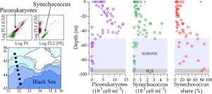
Structure and functions of picophytoplankton and their links to the water column density stratification and nutrient vertical profiles were studied in the Black Sea open waters along a transect from the Western Gyre to the NW shelf during spring homothermy in April of 2017. Abundances of picocyanobacteria of the genus Synechococcus (0.85 ± 0.96 (SD) × 103 cells ml−1) and eukaryotic picoalgae (5.74 ± 5.99 × 10³ cells ml−1) and their intracellular pigment (chl a, phycoerythrin) contents were quantified using flow cytometry. The distribution of these variables in the upper 100-m layer was non-uniform. Along the whole transect, Synechococcus and picoeukaryote abundance maxima (up to 4 × 10³ and 3 × 10⁴ cells ml−1, respectively) were observed at the water temperature of 8–8.5°С in the depth range between 30 and 40 m. Picoeukaryotes dominated the total community. The share of Synecococcus averaged about 22% and increased with depth up to 80%. Picophytoplankton abundance dropped abruptly through the oxycline but the cells were found in the suboxic layer at about 103 cells ml−1 and in a sample from the anoxic zone at 102 cells ml−1. Intracellular chl a maxima were revealed in the nitrate peak at sigma-t density of 15.5. In the surface and suboxic layers, non-specific green autofluorescence (GAF) was detected in picophytoplankton cells that might provide evidence of their stress state in adverse environment. Multivariate analysis has revealed tight coupling between abiotic and microbiological variables in three statistically distinct pelagic regions of the transect, corresponding to the abyss, the continental slope and the shelf.
期刊介绍:
The Journal of Marine Systems provides a medium for interdisciplinary exchange between physical, chemical and biological oceanographers and marine geologists. The journal welcomes original research papers and review articles. Preference will be given to interdisciplinary approaches to marine systems.


 分享
分享
 求助内容:
求助内容: 应助结果提醒方式:
 扫码关注我们
