Hyun-Wook Kim, Seung Hak Oh, Se Jeong Lee, Ji eun Na, Im Joo Rhyu
{"title":"大鼠小脑系统发育小叶中浦肯野细胞树突棘与平行纤维静脉曲张突触密度的差异","authors":"Hyun-Wook Kim, Seung Hak Oh, Se Jeong Lee, Ji eun Na, Im Joo Rhyu","doi":"10.1186/s42649-020-00027-6","DOIUrl":null,"url":null,"abstract":"<p>The cerebellum is a region of the brain that plays an important role in motor control. It is classified phylogenetically into archicerebellum, paleocerebellum and neocerebellum. The Purkinje cells are lined in a row called Purkinje cell layer and it has a unique dendritic branches with many spines.</p><p>The previous study reported that there is a difference of synapse density according to the lobules based on large two-dimensional data. However, recent study with high voltage electron microscopy showed there was no differences in dendritic spine density of the Purkinje cell according to its phylogenetic lobule. We analyzed Purkinje cell density in the II, VI and X lobules by stereological modules and synaptic density was estimated by double disector based on Purkinje cell density in the molecular layer of each lobule.</p><p>The results showed that there was significant difference in the Purkinje cell density and synapse number according to their phylogenetic lobules. The number of Purkinje cell in a given volume was larger in the archicerebellum, but synapse density was higher in the neocerebellum.</p><p>These data suggest that cellular and synaptic organization of the Purkinje cell is different according to their phylogenetic background.</p>","PeriodicalId":470,"journal":{"name":"Applied Microscopy","volume":"50 1","pages":""},"PeriodicalIF":0.0000,"publicationDate":"2020-02-27","publicationTypes":"Journal Article","fieldsOfStudy":null,"isOpenAccess":false,"openAccessPdf":"https://sci-hub-pdf.com/10.1186/s42649-020-00027-6","citationCount":"2","resultStr":"{\"title\":\"Differential synapse density between Purkinje cell dendritic spine and parallel fiber varicosity in the rat cerebellum among the phylogenic lobules\",\"authors\":\"Hyun-Wook Kim, Seung Hak Oh, Se Jeong Lee, Ji eun Na, Im Joo Rhyu\",\"doi\":\"10.1186/s42649-020-00027-6\",\"DOIUrl\":null,\"url\":null,\"abstract\":\"<p>The cerebellum is a region of the brain that plays an important role in motor control. It is classified phylogenetically into archicerebellum, paleocerebellum and neocerebellum. The Purkinje cells are lined in a row called Purkinje cell layer and it has a unique dendritic branches with many spines.</p><p>The previous study reported that there is a difference of synapse density according to the lobules based on large two-dimensional data. However, recent study with high voltage electron microscopy showed there was no differences in dendritic spine density of the Purkinje cell according to its phylogenetic lobule. We analyzed Purkinje cell density in the II, VI and X lobules by stereological modules and synaptic density was estimated by double disector based on Purkinje cell density in the molecular layer of each lobule.</p><p>The results showed that there was significant difference in the Purkinje cell density and synapse number according to their phylogenetic lobules. The number of Purkinje cell in a given volume was larger in the archicerebellum, but synapse density was higher in the neocerebellum.</p><p>These data suggest that cellular and synaptic organization of the Purkinje cell is different according to their phylogenetic background.</p>\",\"PeriodicalId\":470,\"journal\":{\"name\":\"Applied Microscopy\",\"volume\":\"50 1\",\"pages\":\"\"},\"PeriodicalIF\":0.0000,\"publicationDate\":\"2020-02-27\",\"publicationTypes\":\"Journal Article\",\"fieldsOfStudy\":null,\"isOpenAccess\":false,\"openAccessPdf\":\"https://sci-hub-pdf.com/10.1186/s42649-020-00027-6\",\"citationCount\":\"2\",\"resultStr\":null,\"platform\":\"Semanticscholar\",\"paperid\":null,\"PeriodicalName\":\"Applied Microscopy\",\"FirstCategoryId\":\"1085\",\"ListUrlMain\":\"https://link.springer.com/article/10.1186/s42649-020-00027-6\",\"RegionNum\":0,\"RegionCategory\":null,\"ArticlePicture\":[],\"TitleCN\":null,\"AbstractTextCN\":null,\"PMCID\":null,\"EPubDate\":\"\",\"PubModel\":\"\",\"JCR\":\"Q3\",\"JCRName\":\"Immunology and Microbiology\",\"Score\":null,\"Total\":0}","platform":"Semanticscholar","paperid":null,"PeriodicalName":"Applied Microscopy","FirstCategoryId":"1085","ListUrlMain":"https://link.springer.com/article/10.1186/s42649-020-00027-6","RegionNum":0,"RegionCategory":null,"ArticlePicture":[],"TitleCN":null,"AbstractTextCN":null,"PMCID":null,"EPubDate":"","PubModel":"","JCR":"Q3","JCRName":"Immunology and Microbiology","Score":null,"Total":0}
Differential synapse density between Purkinje cell dendritic spine and parallel fiber varicosity in the rat cerebellum among the phylogenic lobules
The cerebellum is a region of the brain that plays an important role in motor control. It is classified phylogenetically into archicerebellum, paleocerebellum and neocerebellum. The Purkinje cells are lined in a row called Purkinje cell layer and it has a unique dendritic branches with many spines.
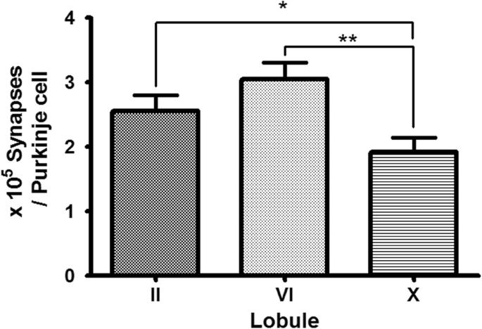
The previous study reported that there is a difference of synapse density according to the lobules based on large two-dimensional data. However, recent study with high voltage electron microscopy showed there was no differences in dendritic spine density of the Purkinje cell according to its phylogenetic lobule. We analyzed Purkinje cell density in the II, VI and X lobules by stereological modules and synaptic density was estimated by double disector based on Purkinje cell density in the molecular layer of each lobule.
The results showed that there was significant difference in the Purkinje cell density and synapse number according to their phylogenetic lobules. The number of Purkinje cell in a given volume was larger in the archicerebellum, but synapse density was higher in the neocerebellum.
These data suggest that cellular and synaptic organization of the Purkinje cell is different according to their phylogenetic background.
Applied MicroscopyImmunology and Microbiology-Applied Microbiology and Biotechnology
CiteScore
3.40
自引率
0.00%
发文量
10
审稿时长
10 weeks
期刊介绍:
Applied Microscopy is a peer-reviewed journal sponsored by the Korean Society of Microscopy. The journal covers all the interdisciplinary fields of technological developments in new microscopy methods and instrumentation and their applications to biological or materials science for determining structure and chemistry. ISSN: 22875123, 22874445.


 分享
分享
 求助内容:
求助内容: 应助结果提醒方式:
 扫码关注我们
