Lucas Glaucio da Silva, Waleska Rayanne Sizinia da Silva Monteiro, Tiago Medeiros de Aguiar Moreira, Maria Aparecida Esteves Rabelo, Emílio Augusto Campos Pereira de Assis, Gustavo Torres de Souza
{"title":"分形维数分析作为一种简便的计算方法来提高乳腺癌的组织病理学诊断","authors":"Lucas Glaucio da Silva, Waleska Rayanne Sizinia da Silva Monteiro, Tiago Medeiros de Aguiar Moreira, Maria Aparecida Esteves Rabelo, Emílio Augusto Campos Pereira de Assis, Gustavo Torres de Souza","doi":"10.1186/s42649-021-00055-w","DOIUrl":null,"url":null,"abstract":"<p>Histopathology is a well-established standard diagnosis employed for the majority of malignancies, including breast cancer. Nevertheless, despite training and standardization, it is considered operator-dependent and errors are still a concern. Fractal dimension analysis is a computational image processing technique that allows assessing the degree of complexity in patterns. We aimed here at providing a robust and easily attainable method for introducing computer-assisted techniques to histopathology laboratories. Slides from two databases were used: A) Breast Cancer Histopathological; and B) Grand Challenge on Breast Cancer Histology. Set A contained 2480 images from 24 patients with benign alterations, and 5429 images from 58 patients with breast cancer. Set B comprised 100 images of each type: normal tissue, benign alterations, in situ carcinoma, and invasive carcinoma. All images were analyzed with the FracLac algorithm in the ImageJ computational environment to yield the box count fractal dimension (Db) results. Images on set A on 40x magnification were statistically different (<i>p</i>?=?0.0003), whereas images on 400x did not present differences in their means. On set B, the mean Db values presented promissing statistical differences when comparing. Normal and/or benign images to in situ and/or invasive carcinoma (all <i>p</i>?<?0.0001). Interestingly, there was no difference when comparing normal tissue to benign alterations. These data corroborate with previous work in which fractal analysis allowed differentiating malignancies. Computer-aided diagnosis algorithms may beneficiate from using Db data; specific Db cut-off values may yield ~?99% specificity in diagnosing breast cancer. Furthermore, the fact that it allows assessing tissue complexity, this tool may be used to understand the progression of the histological alterations in cancer.</p>","PeriodicalId":470,"journal":{"name":"Applied Microscopy","volume":"51 1","pages":""},"PeriodicalIF":0.0000,"publicationDate":"2021-04-30","publicationTypes":"Journal Article","fieldsOfStudy":null,"isOpenAccess":false,"openAccessPdf":"https://sci-hub-pdf.com/10.1186/s42649-021-00055-w","citationCount":"8","resultStr":"{\"title\":\"Fractal dimension analysis as an easy computational approach to improve breast cancer histopathological diagnosis\",\"authors\":\"Lucas Glaucio da Silva, Waleska Rayanne Sizinia da Silva Monteiro, Tiago Medeiros de Aguiar Moreira, Maria Aparecida Esteves Rabelo, Emílio Augusto Campos Pereira de Assis, Gustavo Torres de Souza\",\"doi\":\"10.1186/s42649-021-00055-w\",\"DOIUrl\":null,\"url\":null,\"abstract\":\"<p>Histopathology is a well-established standard diagnosis employed for the majority of malignancies, including breast cancer. Nevertheless, despite training and standardization, it is considered operator-dependent and errors are still a concern. Fractal dimension analysis is a computational image processing technique that allows assessing the degree of complexity in patterns. We aimed here at providing a robust and easily attainable method for introducing computer-assisted techniques to histopathology laboratories. Slides from two databases were used: A) Breast Cancer Histopathological; and B) Grand Challenge on Breast Cancer Histology. Set A contained 2480 images from 24 patients with benign alterations, and 5429 images from 58 patients with breast cancer. Set B comprised 100 images of each type: normal tissue, benign alterations, in situ carcinoma, and invasive carcinoma. All images were analyzed with the FracLac algorithm in the ImageJ computational environment to yield the box count fractal dimension (Db) results. Images on set A on 40x magnification were statistically different (<i>p</i>?=?0.0003), whereas images on 400x did not present differences in their means. On set B, the mean Db values presented promissing statistical differences when comparing. Normal and/or benign images to in situ and/or invasive carcinoma (all <i>p</i>?<?0.0001). Interestingly, there was no difference when comparing normal tissue to benign alterations. These data corroborate with previous work in which fractal analysis allowed differentiating malignancies. Computer-aided diagnosis algorithms may beneficiate from using Db data; specific Db cut-off values may yield ~?99% specificity in diagnosing breast cancer. Furthermore, the fact that it allows assessing tissue complexity, this tool may be used to understand the progression of the histological alterations in cancer.</p>\",\"PeriodicalId\":470,\"journal\":{\"name\":\"Applied Microscopy\",\"volume\":\"51 1\",\"pages\":\"\"},\"PeriodicalIF\":0.0000,\"publicationDate\":\"2021-04-30\",\"publicationTypes\":\"Journal Article\",\"fieldsOfStudy\":null,\"isOpenAccess\":false,\"openAccessPdf\":\"https://sci-hub-pdf.com/10.1186/s42649-021-00055-w\",\"citationCount\":\"8\",\"resultStr\":null,\"platform\":\"Semanticscholar\",\"paperid\":null,\"PeriodicalName\":\"Applied Microscopy\",\"FirstCategoryId\":\"1085\",\"ListUrlMain\":\"https://link.springer.com/article/10.1186/s42649-021-00055-w\",\"RegionNum\":0,\"RegionCategory\":null,\"ArticlePicture\":[],\"TitleCN\":null,\"AbstractTextCN\":null,\"PMCID\":null,\"EPubDate\":\"\",\"PubModel\":\"\",\"JCR\":\"Q3\",\"JCRName\":\"Immunology and Microbiology\",\"Score\":null,\"Total\":0}","platform":"Semanticscholar","paperid":null,"PeriodicalName":"Applied Microscopy","FirstCategoryId":"1085","ListUrlMain":"https://link.springer.com/article/10.1186/s42649-021-00055-w","RegionNum":0,"RegionCategory":null,"ArticlePicture":[],"TitleCN":null,"AbstractTextCN":null,"PMCID":null,"EPubDate":"","PubModel":"","JCR":"Q3","JCRName":"Immunology and Microbiology","Score":null,"Total":0}
Fractal dimension analysis as an easy computational approach to improve breast cancer histopathological diagnosis
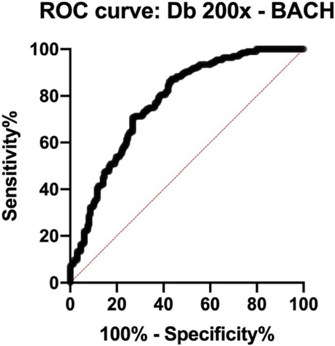
Histopathology is a well-established standard diagnosis employed for the majority of malignancies, including breast cancer. Nevertheless, despite training and standardization, it is considered operator-dependent and errors are still a concern. Fractal dimension analysis is a computational image processing technique that allows assessing the degree of complexity in patterns. We aimed here at providing a robust and easily attainable method for introducing computer-assisted techniques to histopathology laboratories. Slides from two databases were used: A) Breast Cancer Histopathological; and B) Grand Challenge on Breast Cancer Histology. Set A contained 2480 images from 24 patients with benign alterations, and 5429 images from 58 patients with breast cancer. Set B comprised 100 images of each type: normal tissue, benign alterations, in situ carcinoma, and invasive carcinoma. All images were analyzed with the FracLac algorithm in the ImageJ computational environment to yield the box count fractal dimension (Db) results. Images on set A on 40x magnification were statistically different (p?=?0.0003), whereas images on 400x did not present differences in their means. On set B, the mean Db values presented promissing statistical differences when comparing. Normal and/or benign images to in situ and/or invasive carcinoma (all p?<?0.0001). Interestingly, there was no difference when comparing normal tissue to benign alterations. These data corroborate with previous work in which fractal analysis allowed differentiating malignancies. Computer-aided diagnosis algorithms may beneficiate from using Db data; specific Db cut-off values may yield ~?99% specificity in diagnosing breast cancer. Furthermore, the fact that it allows assessing tissue complexity, this tool may be used to understand the progression of the histological alterations in cancer.
Applied MicroscopyImmunology and Microbiology-Applied Microbiology and Biotechnology
CiteScore
3.40
自引率
0.00%
发文量
10
审稿时长
10 weeks
期刊介绍:
Applied Microscopy is a peer-reviewed journal sponsored by the Korean Society of Microscopy. The journal covers all the interdisciplinary fields of technological developments in new microscopy methods and instrumentation and their applications to biological or materials science for determining structure and chemistry. ISSN: 22875123, 22874445.


 分享
分享
 求助内容:
求助内容: 应助结果提醒方式:
 扫码关注我们
