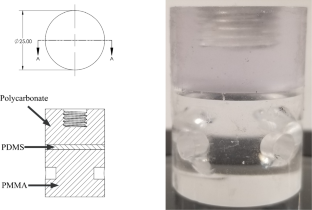
{"title":"弹性体裂纹形核和扩展:1 .原位光学和x射线实验观察","authors":"Jinlong Guo, Krishnaswamy Ravi-Chandar","doi":"10.1007/s10704-023-00714-x","DOIUrl":null,"url":null,"abstract":"<div><p>This article presents the results of an investigation of crack nucleation and propagation in a transparent polydimenthylsiloxane (PDMS) elastomer. The main objective of the investigation is to characterize quantitatively the evolution of crack nucleation and propagation behavior not just through the usual macroscopic load and displacement data, but with synchronized optical images at high spatial and adequate temporal resolution that will resolve the evolution of the failure processes. This is augmented with X-ray computed tomography (CT) scans to characterize the three-dimensional geometry of the cracks nucleated in the interior of the elastomer. Towards this goal, we reproduce the classical <i>poker-chip</i> experiment of Gent and Lindley (Proc R Soc Lond A 249(1257):195–205, 1959) in which the specimen’s diameter-to-thickness ratio is varied over a broad range to cover crack nucleation, propagation, and their coalescence. These experiments are performed on transparent PDMS with different compositions, first in a specially built loading machine that is fitted with a high magnification microscopic camera that permits the measurement of the load while simultaneously providing images of the specimen configuration and subsequently in an apparatus built for in situ observations using an X-ray CT scanning system. These experiments reveal that nucleation of multiple microcracks dominates when the diameter-to-thickness aspect ratio <span>\\(\\alpha \\)</span> is sufficiently large, because the incompressibility of the material induces substantial, nearly uniform hydrostatic tension in the specimen. In contrast, specimens with smaller aspect ratio tend to nucleate fewer cracks, and are dominated by the growth of these cracks. At even smaller <span>\\(\\alpha \\)</span>, the hydrostatic stress is significantly lowered and failure is dominated by surface flaws. The three-dimensional geometry, and the spatial distribution of the nucleated cracks were evaluated using optical microscopy and X-ray CT scans. This revealed cracks of three different shapes, one of which was confined in a layer near to the upper or bottom boundary of the poker-chip, another was across the thickness, but with a tilt relative to the axis of the specimen, and the last was propagating along the radial direction.</p></div>","PeriodicalId":590,"journal":{"name":"International Journal of Fracture","volume":"243 1","pages":"1 - 29"},"PeriodicalIF":2.5000,"publicationDate":"2023-07-27","publicationTypes":"Journal Article","fieldsOfStudy":null,"isOpenAccess":false,"openAccessPdf":"https://link.springer.com/content/pdf/10.1007/s10704-023-00714-x.pdf","citationCount":"0","resultStr":"{\"title\":\"On crack nucleation and propagation in elastomers: I. In situ optical and X-ray experimental observations\",\"authors\":\"Jinlong Guo, Krishnaswamy Ravi-Chandar\",\"doi\":\"10.1007/s10704-023-00714-x\",\"DOIUrl\":null,\"url\":null,\"abstract\":\"<div><p>This article presents the results of an investigation of crack nucleation and propagation in a transparent polydimenthylsiloxane (PDMS) elastomer. The main objective of the investigation is to characterize quantitatively the evolution of crack nucleation and propagation behavior not just through the usual macroscopic load and displacement data, but with synchronized optical images at high spatial and adequate temporal resolution that will resolve the evolution of the failure processes. This is augmented with X-ray computed tomography (CT) scans to characterize the three-dimensional geometry of the cracks nucleated in the interior of the elastomer. Towards this goal, we reproduce the classical <i>poker-chip</i> experiment of Gent and Lindley (Proc R Soc Lond A 249(1257):195–205, 1959) in which the specimen’s diameter-to-thickness ratio is varied over a broad range to cover crack nucleation, propagation, and their coalescence. These experiments are performed on transparent PDMS with different compositions, first in a specially built loading machine that is fitted with a high magnification microscopic camera that permits the measurement of the load while simultaneously providing images of the specimen configuration and subsequently in an apparatus built for in situ observations using an X-ray CT scanning system. These experiments reveal that nucleation of multiple microcracks dominates when the diameter-to-thickness aspect ratio <span>\\\\(\\\\alpha \\\\)</span> is sufficiently large, because the incompressibility of the material induces substantial, nearly uniform hydrostatic tension in the specimen. In contrast, specimens with smaller aspect ratio tend to nucleate fewer cracks, and are dominated by the growth of these cracks. At even smaller <span>\\\\(\\\\alpha \\\\)</span>, the hydrostatic stress is significantly lowered and failure is dominated by surface flaws. The three-dimensional geometry, and the spatial distribution of the nucleated cracks were evaluated using optical microscopy and X-ray CT scans. This revealed cracks of three different shapes, one of which was confined in a layer near to the upper or bottom boundary of the poker-chip, another was across the thickness, but with a tilt relative to the axis of the specimen, and the last was propagating along the radial direction.</p></div>\",\"PeriodicalId\":590,\"journal\":{\"name\":\"International Journal of Fracture\",\"volume\":\"243 1\",\"pages\":\"1 - 29\"},\"PeriodicalIF\":2.5000,\"publicationDate\":\"2023-07-27\",\"publicationTypes\":\"Journal Article\",\"fieldsOfStudy\":null,\"isOpenAccess\":false,\"openAccessPdf\":\"https://link.springer.com/content/pdf/10.1007/s10704-023-00714-x.pdf\",\"citationCount\":\"0\",\"resultStr\":null,\"platform\":\"Semanticscholar\",\"paperid\":null,\"PeriodicalName\":\"International Journal of Fracture\",\"FirstCategoryId\":\"5\",\"ListUrlMain\":\"https://link.springer.com/article/10.1007/s10704-023-00714-x\",\"RegionNum\":3,\"RegionCategory\":\"工程技术\",\"ArticlePicture\":[],\"TitleCN\":null,\"AbstractTextCN\":null,\"PMCID\":null,\"EPubDate\":\"\",\"PubModel\":\"\",\"JCR\":\"Q3\",\"JCRName\":\"MATERIALS SCIENCE, MULTIDISCIPLINARY\",\"Score\":null,\"Total\":0}","platform":"Semanticscholar","paperid":null,"PeriodicalName":"International Journal of Fracture","FirstCategoryId":"5","ListUrlMain":"https://link.springer.com/article/10.1007/s10704-023-00714-x","RegionNum":3,"RegionCategory":"工程技术","ArticlePicture":[],"TitleCN":null,"AbstractTextCN":null,"PMCID":null,"EPubDate":"","PubModel":"","JCR":"Q3","JCRName":"MATERIALS SCIENCE, MULTIDISCIPLINARY","Score":null,"Total":0}
引用次数: 0
摘要
本文介绍了透明聚硅氧烷(PDMS)弹性体裂纹形核和扩展的研究结果。研究的主要目的是定量表征裂纹形核和扩展行为的演变,而不仅仅是通过通常的宏观载荷和位移数据,而是通过高空间和足够的时间分辨率的同步光学图像来解决破坏过程的演变。通过x射线计算机断层扫描(CT),可以对弹性体内部裂缝的三维几何形状进行表征。为了实现这一目标,我们重现了Gent和Lindley的经典扑克片实验(Proc R Soc load A 249(1257):195 - 205,1959),其中试样的直径与厚度比在很宽的范围内变化,以覆盖裂纹的成核、扩展和合并。这些实验是在不同成分的透明PDMS上进行的,首先在一个专门建造的装载机器中进行,该机器配备了一个高放大显微镜相机,可以在测量负载的同时提供样品结构的图像,然后在一个使用x射线CT扫描系统进行现场观察的设备中进行。这些实验表明,当径厚比\(\alpha \)足够大时,多个微裂纹的成核占主导地位,因为材料的不可压缩性在试样中引起了大量的、几乎均匀的静水张力。而纵横比越小的试样,裂纹的形核越少,且以裂纹的扩展为主导。在更小的\(\alpha \),静水应力显著降低,破坏主要是表面缺陷。利用光学显微镜和x射线CT扫描评估了成核裂纹的三维几何形状和空间分布。这揭示了三种不同形状的裂缝,其中一种被限制在靠近扑克片的上或下边界的一层,另一种跨越厚度,但相对于试样的轴线有倾斜,最后一种沿着径向传播。
On crack nucleation and propagation in elastomers: I. In situ optical and X-ray experimental observations
This article presents the results of an investigation of crack nucleation and propagation in a transparent polydimenthylsiloxane (PDMS) elastomer. The main objective of the investigation is to characterize quantitatively the evolution of crack nucleation and propagation behavior not just through the usual macroscopic load and displacement data, but with synchronized optical images at high spatial and adequate temporal resolution that will resolve the evolution of the failure processes. This is augmented with X-ray computed tomography (CT) scans to characterize the three-dimensional geometry of the cracks nucleated in the interior of the elastomer. Towards this goal, we reproduce the classical poker-chip experiment of Gent and Lindley (Proc R Soc Lond A 249(1257):195–205, 1959) in which the specimen’s diameter-to-thickness ratio is varied over a broad range to cover crack nucleation, propagation, and their coalescence. These experiments are performed on transparent PDMS with different compositions, first in a specially built loading machine that is fitted with a high magnification microscopic camera that permits the measurement of the load while simultaneously providing images of the specimen configuration and subsequently in an apparatus built for in situ observations using an X-ray CT scanning system. These experiments reveal that nucleation of multiple microcracks dominates when the diameter-to-thickness aspect ratio \(\alpha \) is sufficiently large, because the incompressibility of the material induces substantial, nearly uniform hydrostatic tension in the specimen. In contrast, specimens with smaller aspect ratio tend to nucleate fewer cracks, and are dominated by the growth of these cracks. At even smaller \(\alpha \), the hydrostatic stress is significantly lowered and failure is dominated by surface flaws. The three-dimensional geometry, and the spatial distribution of the nucleated cracks were evaluated using optical microscopy and X-ray CT scans. This revealed cracks of three different shapes, one of which was confined in a layer near to the upper or bottom boundary of the poker-chip, another was across the thickness, but with a tilt relative to the axis of the specimen, and the last was propagating along the radial direction.
期刊介绍:
The International Journal of Fracture is an outlet for original analytical, numerical and experimental contributions which provide improved understanding of the mechanisms of micro and macro fracture in all materials, and their engineering implications.
The Journal is pleased to receive papers from engineers and scientists working in various aspects of fracture. Contributions emphasizing empirical correlations, unanalyzed experimental results or routine numerical computations, while representing important necessary aspects of certain fatigue, strength, and fracture analyses, will normally be discouraged; occasional review papers in these as well as other areas are welcomed. Innovative and in-depth engineering applications of fracture theory are also encouraged.
In addition, the Journal welcomes, for rapid publication, Brief Notes in Fracture and Micromechanics which serve the Journal''s Objective. Brief Notes include: Brief presentation of a new idea, concept or method; new experimental observations or methods of significance; short notes of quality that do not amount to full length papers; discussion of previously published work in the Journal, and Brief Notes Errata.


 分享
分享
 求助内容:
求助内容: 应助结果提醒方式:
 扫码关注我们
