{"title":"翼管形态和地形的变异分析及其对神经外科的影响:骨测量学研究。","authors":"Navita Aggarwal, Noopinder Kaur, Apurba Patra, Monika Gupta","doi":"10.1055/s-0043-1772759","DOIUrl":null,"url":null,"abstract":"<p><p><b>Objective</b> Pterion is an \"H\" shaped formation of sutures located in the temporal fossa of the skull. It is an important anatomical landmark and a craniometric point. The thinness of the skull and its inner relation with the middle meningeal artery make this anatomical landmark clinically significant. Variations in the pterion are imperative, especially for neurosurgeons in order to have the most suitable craniometric point to be minimally invasive. <b>Materials and Methods</b> One hundred pterions were studied to report the variations in the type and location of the pterion. Murphy's classification was used to classify the pterion into four types on the basis of bone articulation-sphenoparietal, frontotemporal, stellate, and epipteric. <b>Results</b> All four types of pterions were observed, sphenoparietal being the most common. No significant gender difference was observed in terms of type and laterality of various pterions. The mean distance between the center of pterion to the superolateral point of zygomaticotemporal (PZT) suture and the anterolateral point of the frontozygomatic (PFZ) suture were 3.91 ± 3.79 cm and 3.68 ± 3.79 mm, respectively. Correlation analysis showed a strong positive relation between PZT and PFZ sutures. <b>Conclusion</b> Accurate data on the morphology and morphometry of bony anatomical points are crucial, while performing intracranial surgery using them as recognizable landmarks. The morphometric parameters may help in determining the soundness of the pterion as an identifiable landmark for performing interventions like burr hole and other neurosurgical procedures in this area.</p>","PeriodicalId":6888,"journal":{"name":"Acta Histochemica Et Cytochemica","volume":"22 1","pages":"581-586"},"PeriodicalIF":1.9000,"publicationDate":"2023-08-31","publicationTypes":"Journal Article","fieldsOfStudy":null,"isOpenAccess":false,"openAccessPdf":"https://www.ncbi.nlm.nih.gov/pmc/articles/PMC10749850/pdf/","citationCount":"0","resultStr":"{\"title\":\"Analysis of the Variations in the Morphology, Topography of the Pterion, and Their Implications in Neurosurgery: An Osteometric Study.\",\"authors\":\"Navita Aggarwal, Noopinder Kaur, Apurba Patra, Monika Gupta\",\"doi\":\"10.1055/s-0043-1772759\",\"DOIUrl\":null,\"url\":null,\"abstract\":\"<p><p><b>Objective</b> Pterion is an \\\"H\\\" shaped formation of sutures located in the temporal fossa of the skull. It is an important anatomical landmark and a craniometric point. The thinness of the skull and its inner relation with the middle meningeal artery make this anatomical landmark clinically significant. Variations in the pterion are imperative, especially for neurosurgeons in order to have the most suitable craniometric point to be minimally invasive. <b>Materials and Methods</b> One hundred pterions were studied to report the variations in the type and location of the pterion. Murphy's classification was used to classify the pterion into four types on the basis of bone articulation-sphenoparietal, frontotemporal, stellate, and epipteric. <b>Results</b> All four types of pterions were observed, sphenoparietal being the most common. No significant gender difference was observed in terms of type and laterality of various pterions. The mean distance between the center of pterion to the superolateral point of zygomaticotemporal (PZT) suture and the anterolateral point of the frontozygomatic (PFZ) suture were 3.91 ± 3.79 cm and 3.68 ± 3.79 mm, respectively. Correlation analysis showed a strong positive relation between PZT and PFZ sutures. <b>Conclusion</b> Accurate data on the morphology and morphometry of bony anatomical points are crucial, while performing intracranial surgery using them as recognizable landmarks. The morphometric parameters may help in determining the soundness of the pterion as an identifiable landmark for performing interventions like burr hole and other neurosurgical procedures in this area.</p>\",\"PeriodicalId\":6888,\"journal\":{\"name\":\"Acta Histochemica Et Cytochemica\",\"volume\":\"22 1\",\"pages\":\"581-586\"},\"PeriodicalIF\":1.9000,\"publicationDate\":\"2023-08-31\",\"publicationTypes\":\"Journal Article\",\"fieldsOfStudy\":null,\"isOpenAccess\":false,\"openAccessPdf\":\"https://www.ncbi.nlm.nih.gov/pmc/articles/PMC10749850/pdf/\",\"citationCount\":\"0\",\"resultStr\":null,\"platform\":\"Semanticscholar\",\"paperid\":null,\"PeriodicalName\":\"Acta Histochemica Et Cytochemica\",\"FirstCategoryId\":\"1085\",\"ListUrlMain\":\"https://doi.org/10.1055/s-0043-1772759\",\"RegionNum\":4,\"RegionCategory\":\"生物学\",\"ArticlePicture\":[],\"TitleCN\":null,\"AbstractTextCN\":null,\"PMCID\":null,\"EPubDate\":\"2023/9/1 0:00:00\",\"PubModel\":\"eCollection\",\"JCR\":\"Q4\",\"JCRName\":\"CELL BIOLOGY\",\"Score\":null,\"Total\":0}","platform":"Semanticscholar","paperid":null,"PeriodicalName":"Acta Histochemica Et Cytochemica","FirstCategoryId":"1085","ListUrlMain":"https://doi.org/10.1055/s-0043-1772759","RegionNum":4,"RegionCategory":"生物学","ArticlePicture":[],"TitleCN":null,"AbstractTextCN":null,"PMCID":null,"EPubDate":"2023/9/1 0:00:00","PubModel":"eCollection","JCR":"Q4","JCRName":"CELL BIOLOGY","Score":null,"Total":0}
Analysis of the Variations in the Morphology, Topography of the Pterion, and Their Implications in Neurosurgery: An Osteometric Study.
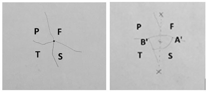
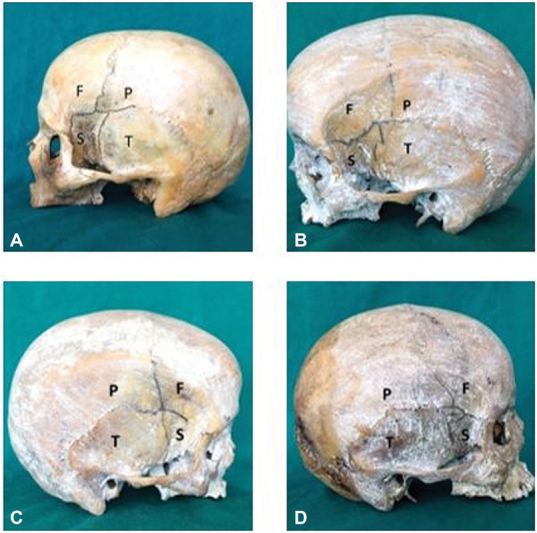
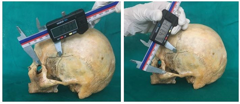
Objective Pterion is an "H" shaped formation of sutures located in the temporal fossa of the skull. It is an important anatomical landmark and a craniometric point. The thinness of the skull and its inner relation with the middle meningeal artery make this anatomical landmark clinically significant. Variations in the pterion are imperative, especially for neurosurgeons in order to have the most suitable craniometric point to be minimally invasive. Materials and Methods One hundred pterions were studied to report the variations in the type and location of the pterion. Murphy's classification was used to classify the pterion into four types on the basis of bone articulation-sphenoparietal, frontotemporal, stellate, and epipteric. Results All four types of pterions were observed, sphenoparietal being the most common. No significant gender difference was observed in terms of type and laterality of various pterions. The mean distance between the center of pterion to the superolateral point of zygomaticotemporal (PZT) suture and the anterolateral point of the frontozygomatic (PFZ) suture were 3.91 ± 3.79 cm and 3.68 ± 3.79 mm, respectively. Correlation analysis showed a strong positive relation between PZT and PFZ sutures. Conclusion Accurate data on the morphology and morphometry of bony anatomical points are crucial, while performing intracranial surgery using them as recognizable landmarks. The morphometric parameters may help in determining the soundness of the pterion as an identifiable landmark for performing interventions like burr hole and other neurosurgical procedures in this area.
期刊介绍:
Acta Histochemica et Cytochemica is the official online journal of the Japan Society of Histochemistry and Cytochemistry. It is intended primarily for rapid publication of concise, original articles in the fields of histochemistry and cytochemistry. Manuscripts oriented towards methodological subjects that contain significant technical advances in these fields are also welcome. Manuscripts in English are accepted from investigators in any country, whether or not they are members of the Japan Society of Histochemistry and Cytochemistry. Manuscripts should be original work that has not been previously published and is not being considered for publication elsewhere, with the exception of abstracts. Manuscripts with essentially the same content as a paper that has been published or accepted, or is under consideration for publication, will not be considered. All submitted papers will be peer-reviewed by at least two referees selected by an appropriate Associate Editor. Acceptance is based on scientific significance, originality, and clarity. When required, a revised manuscript should be submitted within 3 months, otherwise it will be considered to be a new submission. The Editor-in-Chief will make all final decisions regarding acceptance.


 分享
分享
 求助内容:
求助内容: 应助结果提醒方式:
 扫码关注我们
