Carlos Alexandre Curylofo Corsi, Claudia Tarcila Gomes Sares, Fabiola Mestriner, Jéssyca Michelon-Barbosa, Vinicius Flora Dugaich, Timna Varela Martins, Alex Martins Násare, Roberta Ribeiro Costa Rosales, Maria Cecília Jordani, José Carlos Alves-Filho, Rodolfo Borges Dos Reis, Mauricio Serra Ribeiro, Christiane Becari
{"title":"从脑死亡供体中分离和原代培养人腹主动脉平滑肌细胞:血管疾病的实验模型。","authors":"Carlos Alexandre Curylofo Corsi, Claudia Tarcila Gomes Sares, Fabiola Mestriner, Jéssyca Michelon-Barbosa, Vinicius Flora Dugaich, Timna Varela Martins, Alex Martins Násare, Roberta Ribeiro Costa Rosales, Maria Cecília Jordani, José Carlos Alves-Filho, Rodolfo Borges Dos Reis, Mauricio Serra Ribeiro, Christiane Becari","doi":"10.1007/s10561-023-10091-3","DOIUrl":null,"url":null,"abstract":"<p><p>Primary cell cultures are essential tools for elucidating the physiopathological mechanisms of the cardiovascular system. Therefore, a primary culture growth protocol of cardiovascular smooth muscle cells (VSMCs) obtained from human abdominal aortas was standardized. Ten abdominal aorta samples were obtained from patients diagnosed with brain death who were organ and tissue donors with family consent. After surgical ablation to capture the aorta, the aortic tissue was removed, immersed in a Custodiol® solution, and kept between 2 and 8 °C. In the laboratory, in a sterile environment, the tissue was fragmented and incubated in culture plates containing an enriched culture medium (DMEM/G/10% fetal bovine serum, L-glutamine, antibiotics and antifungals) and kept in an oven at 37 °C and 5% CO<sub>2</sub>. The aorta was removed after 24 h of incubation, and the culture medium was changed every six days for twenty days. Cell growth was confirmed through morphological analysis using an inverted optical microscope (Nikon®) and immunofluorescence for smooth muscle alpha-actin and nuclei. The development of the VSMCs was observed, and from the twelfth day, differentiation, long cytoplasmic projections, and adjacent cell connections occurred. On the twentieth day, the morphology of the VSMCs was confirmed by actin fiber immunofluorescence, which is a typical characteristic of VSMCs. The standardization allowed VSMC growth and the replicability of the in vitro test, providing a protocol that mimics natural physiological environments for a better understanding of the cardiovascular system. Its use is intended for investigation, tissue bioengineering, and pharmacological treatments.</p>","PeriodicalId":9723,"journal":{"name":"Cell and Tissue Banking","volume":" ","pages":"187-194"},"PeriodicalIF":1.4000,"publicationDate":"2024-03-01","publicationTypes":"Journal Article","fieldsOfStudy":null,"isOpenAccess":false,"openAccessPdf":"","citationCount":"0","resultStr":"{\"title\":\"Isolation and primary culture of human abdominal aorta smooth muscle cells from brain-dead donors: an experimental model for vascular diseases.\",\"authors\":\"Carlos Alexandre Curylofo Corsi, Claudia Tarcila Gomes Sares, Fabiola Mestriner, Jéssyca Michelon-Barbosa, Vinicius Flora Dugaich, Timna Varela Martins, Alex Martins Násare, Roberta Ribeiro Costa Rosales, Maria Cecília Jordani, José Carlos Alves-Filho, Rodolfo Borges Dos Reis, Mauricio Serra Ribeiro, Christiane Becari\",\"doi\":\"10.1007/s10561-023-10091-3\",\"DOIUrl\":null,\"url\":null,\"abstract\":\"<p><p>Primary cell cultures are essential tools for elucidating the physiopathological mechanisms of the cardiovascular system. Therefore, a primary culture growth protocol of cardiovascular smooth muscle cells (VSMCs) obtained from human abdominal aortas was standardized. Ten abdominal aorta samples were obtained from patients diagnosed with brain death who were organ and tissue donors with family consent. After surgical ablation to capture the aorta, the aortic tissue was removed, immersed in a Custodiol® solution, and kept between 2 and 8 °C. In the laboratory, in a sterile environment, the tissue was fragmented and incubated in culture plates containing an enriched culture medium (DMEM/G/10% fetal bovine serum, L-glutamine, antibiotics and antifungals) and kept in an oven at 37 °C and 5% CO<sub>2</sub>. The aorta was removed after 24 h of incubation, and the culture medium was changed every six days for twenty days. Cell growth was confirmed through morphological analysis using an inverted optical microscope (Nikon®) and immunofluorescence for smooth muscle alpha-actin and nuclei. The development of the VSMCs was observed, and from the twelfth day, differentiation, long cytoplasmic projections, and adjacent cell connections occurred. On the twentieth day, the morphology of the VSMCs was confirmed by actin fiber immunofluorescence, which is a typical characteristic of VSMCs. The standardization allowed VSMC growth and the replicability of the in vitro test, providing a protocol that mimics natural physiological environments for a better understanding of the cardiovascular system. Its use is intended for investigation, tissue bioengineering, and pharmacological treatments.</p>\",\"PeriodicalId\":9723,\"journal\":{\"name\":\"Cell and Tissue Banking\",\"volume\":\" \",\"pages\":\"187-194\"},\"PeriodicalIF\":1.4000,\"publicationDate\":\"2024-03-01\",\"publicationTypes\":\"Journal Article\",\"fieldsOfStudy\":null,\"isOpenAccess\":false,\"openAccessPdf\":\"\",\"citationCount\":\"0\",\"resultStr\":null,\"platform\":\"Semanticscholar\",\"paperid\":null,\"PeriodicalName\":\"Cell and Tissue Banking\",\"FirstCategoryId\":\"5\",\"ListUrlMain\":\"https://doi.org/10.1007/s10561-023-10091-3\",\"RegionNum\":4,\"RegionCategory\":\"医学\",\"ArticlePicture\":[],\"TitleCN\":null,\"AbstractTextCN\":null,\"PMCID\":null,\"EPubDate\":\"2023/5/5 0:00:00\",\"PubModel\":\"Epub\",\"JCR\":\"Q4\",\"JCRName\":\"CELL BIOLOGY\",\"Score\":null,\"Total\":0}","platform":"Semanticscholar","paperid":null,"PeriodicalName":"Cell and Tissue Banking","FirstCategoryId":"5","ListUrlMain":"https://doi.org/10.1007/s10561-023-10091-3","RegionNum":4,"RegionCategory":"医学","ArticlePicture":[],"TitleCN":null,"AbstractTextCN":null,"PMCID":null,"EPubDate":"2023/5/5 0:00:00","PubModel":"Epub","JCR":"Q4","JCRName":"CELL BIOLOGY","Score":null,"Total":0}
Isolation and primary culture of human abdominal aorta smooth muscle cells from brain-dead donors: an experimental model for vascular diseases.
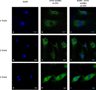
Primary cell cultures are essential tools for elucidating the physiopathological mechanisms of the cardiovascular system. Therefore, a primary culture growth protocol of cardiovascular smooth muscle cells (VSMCs) obtained from human abdominal aortas was standardized. Ten abdominal aorta samples were obtained from patients diagnosed with brain death who were organ and tissue donors with family consent. After surgical ablation to capture the aorta, the aortic tissue was removed, immersed in a Custodiol® solution, and kept between 2 and 8 °C. In the laboratory, in a sterile environment, the tissue was fragmented and incubated in culture plates containing an enriched culture medium (DMEM/G/10% fetal bovine serum, L-glutamine, antibiotics and antifungals) and kept in an oven at 37 °C and 5% CO2. The aorta was removed after 24 h of incubation, and the culture medium was changed every six days for twenty days. Cell growth was confirmed through morphological analysis using an inverted optical microscope (Nikon®) and immunofluorescence for smooth muscle alpha-actin and nuclei. The development of the VSMCs was observed, and from the twelfth day, differentiation, long cytoplasmic projections, and adjacent cell connections occurred. On the twentieth day, the morphology of the VSMCs was confirmed by actin fiber immunofluorescence, which is a typical characteristic of VSMCs. The standardization allowed VSMC growth and the replicability of the in vitro test, providing a protocol that mimics natural physiological environments for a better understanding of the cardiovascular system. Its use is intended for investigation, tissue bioengineering, and pharmacological treatments.
期刊介绍:
Cell and Tissue Banking provides a forum for disseminating information to scientists and clinicians involved in the banking and transplantation of cells and tissues. Cell and Tissue Banking is an international, peer-reviewed journal that publishes original papers in the following areas:
basic research concerning general aspects of tissue banking such as quality assurance and control of banked cells/tissues, effects of preservation and sterilisation methods on cells/tissues, biotechnology, etc.; clinical applications of banked cells/tissues; standards of practice in procurement, processing, storage and distribution of cells/tissues; ethical issues; medico-legal issues.


 分享
分享
 求助内容:
求助内容: 应助结果提醒方式:
 扫码关注我们
