Manzar Ashtari, Mikhail Lipin, Michelle Duong, Gui-Shuang Ying, Yinxi Yu, Albert Maguire, Jean Bennett
{"title":"一组RPE65突变患者视网膜基因治疗对外侧膝状核神经可塑性的影响。","authors":"Manzar Ashtari, Mikhail Lipin, Michelle Duong, Gui-Shuang Ying, Yinxi Yu, Albert Maguire, Jean Bennett","doi":"10.2147/EB.S377275","DOIUrl":null,"url":null,"abstract":"<p><strong>Introduction: </strong>Previous works on experience-dependent brain plasticity have been limited to the cortical structures, overlooking subcortical visual structures such as the lateral geniculate nucleus (LGN). Animal studies have shown substantial experience dependent plasticity and using fMRI, human studies have demonstrated similar properties in patients with cataract surgery. However, in neither animal nor human studies LGN has not been directly assessed, mainly due to its small size, tissue heterogeneity, low contrast/noise ratio, and low spatial resolution.</p><p><strong>Methods: </strong>Utilizing a new algorithm that markedly improves the LGN visibility, LGN was evaluated in a group of low vision patients before and after retinal intervention to reinstate vision and normal sighted matched controls.</p><p><strong>Results: </strong>Between and within groups comparisons showed that patients had significantly smaller left (p< 0.0001) and right (p < 0.00002) LGN volumes at baseline as compared to the one-year follow-up volumes. The same baseline and one year comparison in controls was not significant. Significant positive correlations were observed between the incremental volume increase after gene therapy of the left LGN and the incremental increase in the right (r = 0.71, p < 0.02) and left (r = 0.72, p = 0.018) visual fields. Incremental volume increase of the right LGN also showed a similar positive slope but did not reach significance.</p><p><strong>Discussion: </strong>These results show that despite significantly less volume at baseline, retinal gene therapy promotes robust expansion and increase in LGN volume. Reinstating vision may have facilitated the establishment of new connections between the retina and the LGN and/or unmasking of the dormant connections. The exact trajectory of the structural changes taking place in LGN is unclear but our data shows that even after years of low vision, the LGN in RPE65 patients has the potential for plasticity and expansion to a nearly normal volume one year after gene therapy administration.</p>","PeriodicalId":51844,"journal":{"name":"Eye and Brain","volume":"14 ","pages":"137-147"},"PeriodicalIF":2.4000,"publicationDate":"2022-01-01","publicationTypes":"Journal Article","fieldsOfStudy":null,"isOpenAccess":false,"openAccessPdf":"https://ftp.ncbi.nlm.nih.gov/pub/pmc/oa_pdf/39/70/eb-14-137.PMC9749418.pdf","citationCount":"0","resultStr":"{\"title\":\"Neuroplasticity of the Lateral Geniculate Nucleus in Response to Retinal Gene Therapy in a Group of Patients with <i>RPE65</i> Mutations.\",\"authors\":\"Manzar Ashtari, Mikhail Lipin, Michelle Duong, Gui-Shuang Ying, Yinxi Yu, Albert Maguire, Jean Bennett\",\"doi\":\"10.2147/EB.S377275\",\"DOIUrl\":null,\"url\":null,\"abstract\":\"<p><strong>Introduction: </strong>Previous works on experience-dependent brain plasticity have been limited to the cortical structures, overlooking subcortical visual structures such as the lateral geniculate nucleus (LGN). Animal studies have shown substantial experience dependent plasticity and using fMRI, human studies have demonstrated similar properties in patients with cataract surgery. However, in neither animal nor human studies LGN has not been directly assessed, mainly due to its small size, tissue heterogeneity, low contrast/noise ratio, and low spatial resolution.</p><p><strong>Methods: </strong>Utilizing a new algorithm that markedly improves the LGN visibility, LGN was evaluated in a group of low vision patients before and after retinal intervention to reinstate vision and normal sighted matched controls.</p><p><strong>Results: </strong>Between and within groups comparisons showed that patients had significantly smaller left (p< 0.0001) and right (p < 0.00002) LGN volumes at baseline as compared to the one-year follow-up volumes. The same baseline and one year comparison in controls was not significant. Significant positive correlations were observed between the incremental volume increase after gene therapy of the left LGN and the incremental increase in the right (r = 0.71, p < 0.02) and left (r = 0.72, p = 0.018) visual fields. Incremental volume increase of the right LGN also showed a similar positive slope but did not reach significance.</p><p><strong>Discussion: </strong>These results show that despite significantly less volume at baseline, retinal gene therapy promotes robust expansion and increase in LGN volume. Reinstating vision may have facilitated the establishment of new connections between the retina and the LGN and/or unmasking of the dormant connections. The exact trajectory of the structural changes taking place in LGN is unclear but our data shows that even after years of low vision, the LGN in RPE65 patients has the potential for plasticity and expansion to a nearly normal volume one year after gene therapy administration.</p>\",\"PeriodicalId\":51844,\"journal\":{\"name\":\"Eye and Brain\",\"volume\":\"14 \",\"pages\":\"137-147\"},\"PeriodicalIF\":2.4000,\"publicationDate\":\"2022-01-01\",\"publicationTypes\":\"Journal Article\",\"fieldsOfStudy\":null,\"isOpenAccess\":false,\"openAccessPdf\":\"https://ftp.ncbi.nlm.nih.gov/pub/pmc/oa_pdf/39/70/eb-14-137.PMC9749418.pdf\",\"citationCount\":\"0\",\"resultStr\":null,\"platform\":\"Semanticscholar\",\"paperid\":null,\"PeriodicalName\":\"Eye and Brain\",\"FirstCategoryId\":\"1085\",\"ListUrlMain\":\"https://doi.org/10.2147/EB.S377275\",\"RegionNum\":0,\"RegionCategory\":null,\"ArticlePicture\":[],\"TitleCN\":null,\"AbstractTextCN\":null,\"PMCID\":null,\"EPubDate\":\"\",\"PubModel\":\"\",\"JCR\":\"Q1\",\"JCRName\":\"OPHTHALMOLOGY\",\"Score\":null,\"Total\":0}","platform":"Semanticscholar","paperid":null,"PeriodicalName":"Eye and Brain","FirstCategoryId":"1085","ListUrlMain":"https://doi.org/10.2147/EB.S377275","RegionNum":0,"RegionCategory":null,"ArticlePicture":[],"TitleCN":null,"AbstractTextCN":null,"PMCID":null,"EPubDate":"","PubModel":"","JCR":"Q1","JCRName":"OPHTHALMOLOGY","Score":null,"Total":0}
引用次数: 0
摘要
以往关于经验依赖大脑可塑性的研究仅限于皮层结构,忽视了皮层下的视觉结构,如外侧膝状核(LGN)。动物研究已经显示了大量的经验依赖可塑性,使用功能磁共振成像,人类研究已经在白内障手术患者中证明了类似的特性。然而,在动物和人类研究中,LGN都没有被直接评估,主要是由于其体积小、组织异质性、低对比度/噪声比和低空间分辨率。方法:采用一种明显提高LGN可见度的新算法,对一组低视力患者进行视网膜干预恢复视力前后的LGN进行评估,并与正常视力对照进行比较。结果:组间和组内比较显示,基线时患者左侧LGN体积(p< 0.0001)和右侧LGN体积(p< 0.00002)明显小于1年随访时的体积。相同的基线和对照组的一年比较无显著性。左侧LGN基因治疗后体积增量与右侧视野(r = 0.71, p < 0.02)、左侧视野(r = 0.72, p = 0.018)体积增量呈显著正相关。右侧LGN的增量体积增加也呈现类似的正斜率,但没有达到显著性。讨论:这些结果表明,尽管在基线时体积明显减少,视网膜基因治疗促进了LGN的强劲扩张和体积的增加。恢复视力可能有助于在视网膜和LGN之间建立新的连接和/或揭开休眠连接的面纱。LGN发生结构变化的确切轨迹尚不清楚,但我们的数据显示,即使经过多年的低视力,RPE65患者的LGN在接受基因治疗一年后仍具有可塑性和扩大到接近正常体积的潜力。
Neuroplasticity of the Lateral Geniculate Nucleus in Response to Retinal Gene Therapy in a Group of Patients with RPE65 Mutations.
Introduction: Previous works on experience-dependent brain plasticity have been limited to the cortical structures, overlooking subcortical visual structures such as the lateral geniculate nucleus (LGN). Animal studies have shown substantial experience dependent plasticity and using fMRI, human studies have demonstrated similar properties in patients with cataract surgery. However, in neither animal nor human studies LGN has not been directly assessed, mainly due to its small size, tissue heterogeneity, low contrast/noise ratio, and low spatial resolution.
Methods: Utilizing a new algorithm that markedly improves the LGN visibility, LGN was evaluated in a group of low vision patients before and after retinal intervention to reinstate vision and normal sighted matched controls.
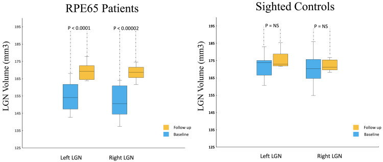
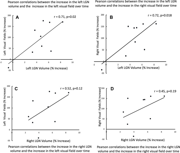
Results: Between and within groups comparisons showed that patients had significantly smaller left (p< 0.0001) and right (p < 0.00002) LGN volumes at baseline as compared to the one-year follow-up volumes. The same baseline and one year comparison in controls was not significant. Significant positive correlations were observed between the incremental volume increase after gene therapy of the left LGN and the incremental increase in the right (r = 0.71, p < 0.02) and left (r = 0.72, p = 0.018) visual fields. Incremental volume increase of the right LGN also showed a similar positive slope but did not reach significance.
Discussion: These results show that despite significantly less volume at baseline, retinal gene therapy promotes robust expansion and increase in LGN volume. Reinstating vision may have facilitated the establishment of new connections between the retina and the LGN and/or unmasking of the dormant connections. The exact trajectory of the structural changes taking place in LGN is unclear but our data shows that even after years of low vision, the LGN in RPE65 patients has the potential for plasticity and expansion to a nearly normal volume one year after gene therapy administration.
期刊介绍:
Eye and Brain is an international, peer-reviewed, open access journal focusing on basic research, clinical findings, and expert reviews in the field of visual science and neuro-ophthalmology. The journal’s unique focus is the link between two well-known visual centres, the eye and the brain, with an emphasis on the importance of such connections. All aspects of clinical and especially basic research on the visual system are addressed within the journal as well as significant future directions in vision research and therapeutic measures. This unique journal focuses on neurological aspects of vision – both physiological and pathological. The scope of the journal spans from the cornea to the associational visual cortex and all the visual centers in between. Topics range from basic biological mechanisms to therapeutic treatment, from simple organisms to humans, and utilizing techniques from molecular biology to behavior. The journal especially welcomes primary research articles or review papers that make the connection between the eye and the brain. Specific areas covered in the journal include: Physiology and pathophysiology of visual centers, Eye movement disorders and strabismus, Cellular, biochemical, and molecular features of the visual system, Structural and functional organization of the eye and of the visual cortex, Metabolic demands of the visual system, Diseases and disorders with neuro-ophthalmic manifestations, Clinical and experimental neuro-ophthalmology and visual system pathologies, Epidemiological studies.


 分享
分享
 求助内容:
求助内容: 应助结果提醒方式:
 扫码关注我们
