{"title":"基于家庭冷冻切片诊断报告的远程数字病理学验证","authors":"Rajiv Kumar Kaushal, Subhash Yadav, Ayushi Sahay, Nupur Karnik, Tushar Agrawal, Vinayak Dave, Nikhil Singh, Ashish Shah, Sangeeta B. Desai","doi":"10.1016/j.jpi.2023.100312","DOIUrl":null,"url":null,"abstract":"<div><h3>Background</h3><p>Despite the promising applications of whole-slide imaging (WSI) for frozen section (FS) diagnosis, its adoption for remote reporting is limited.</p></div><div><h3>Objective</h3><p>To assess the feasibility and performance of home-based remote digital consultation for FS diagnosis.</p></div><div><h3>Material & Method</h3><p>Cases accessioned beyond regular working hours (5 pm–10 pm) were reported simultaneously using optical microscopy (OM) and WSI. Validation of WSI for FS diagnosis from a remote site, i.e. home, was performed by 5 pathologists. Cases were scanned using a portable scanner (Grundium Ocus®40) and previewed on consumer-grade computer devices through a web-based browser (<span>http://grundium.net</span><svg><path></path></svg>). Clinical data and diagnostic reports were shared through a google spreadsheet. The diagnostic concordance, inter- and intra-observer agreement for FS diagnosis by WSI versus OM, and turnaround time (TAT), were recorded.</p></div><div><h3>Results</h3><p>The overall diagnostic accuracy for OM and WSI (from home) was 98.2% (range 97%–100%) and 97.6% (range 95%–99%), respectively, when compared with the reference standard. Almost perfect inter-observer (k = 0.993) and intra-observer (k = 0.987) agreement for WSI was observed by 4 pathologists. Pathologists used consumer-grade laptops/desktops with an average screen size of 14.58 inches (range = 12.3–17.7 inches) and a network speed of 64 megabits per second (range: 10–90 Mbps). The mean diagnostic assessment time per case for OM and WSI was 1:48 min and 5:54 min, respectively. Mean TAT of 27.27 min per case was observed using WSI from home. Seamless connectivity was observed in approximately 75% of cases.</p></div><div><h3>Conclusion</h3><p>This study validates the role of WSI for remote FS diagnosis for its safe and efficient adoption in clinical use.</p></div>","PeriodicalId":37769,"journal":{"name":"Journal of Pathology Informatics","volume":"14 ","pages":"Article 100312"},"PeriodicalIF":0.0000,"publicationDate":"2023-01-01","publicationTypes":"Journal Article","fieldsOfStudy":null,"isOpenAccess":false,"openAccessPdf":"https://ftp.ncbi.nlm.nih.gov/pub/pmc/oa_pdf/81/ac/main.PMC10192998.pdf","citationCount":"0","resultStr":"{\"title\":\"Validation of Remote Digital Pathology based diagnostic reporting of Frozen Sections from home\",\"authors\":\"Rajiv Kumar Kaushal, Subhash Yadav, Ayushi Sahay, Nupur Karnik, Tushar Agrawal, Vinayak Dave, Nikhil Singh, Ashish Shah, Sangeeta B. Desai\",\"doi\":\"10.1016/j.jpi.2023.100312\",\"DOIUrl\":null,\"url\":null,\"abstract\":\"<div><h3>Background</h3><p>Despite the promising applications of whole-slide imaging (WSI) for frozen section (FS) diagnosis, its adoption for remote reporting is limited.</p></div><div><h3>Objective</h3><p>To assess the feasibility and performance of home-based remote digital consultation for FS diagnosis.</p></div><div><h3>Material & Method</h3><p>Cases accessioned beyond regular working hours (5 pm–10 pm) were reported simultaneously using optical microscopy (OM) and WSI. Validation of WSI for FS diagnosis from a remote site, i.e. home, was performed by 5 pathologists. Cases were scanned using a portable scanner (Grundium Ocus®40) and previewed on consumer-grade computer devices through a web-based browser (<span>http://grundium.net</span><svg><path></path></svg>). Clinical data and diagnostic reports were shared through a google spreadsheet. The diagnostic concordance, inter- and intra-observer agreement for FS diagnosis by WSI versus OM, and turnaround time (TAT), were recorded.</p></div><div><h3>Results</h3><p>The overall diagnostic accuracy for OM and WSI (from home) was 98.2% (range 97%–100%) and 97.6% (range 95%–99%), respectively, when compared with the reference standard. Almost perfect inter-observer (k = 0.993) and intra-observer (k = 0.987) agreement for WSI was observed by 4 pathologists. Pathologists used consumer-grade laptops/desktops with an average screen size of 14.58 inches (range = 12.3–17.7 inches) and a network speed of 64 megabits per second (range: 10–90 Mbps). The mean diagnostic assessment time per case for OM and WSI was 1:48 min and 5:54 min, respectively. Mean TAT of 27.27 min per case was observed using WSI from home. Seamless connectivity was observed in approximately 75% of cases.</p></div><div><h3>Conclusion</h3><p>This study validates the role of WSI for remote FS diagnosis for its safe and efficient adoption in clinical use.</p></div>\",\"PeriodicalId\":37769,\"journal\":{\"name\":\"Journal of Pathology Informatics\",\"volume\":\"14 \",\"pages\":\"Article 100312\"},\"PeriodicalIF\":0.0000,\"publicationDate\":\"2023-01-01\",\"publicationTypes\":\"Journal Article\",\"fieldsOfStudy\":null,\"isOpenAccess\":false,\"openAccessPdf\":\"https://ftp.ncbi.nlm.nih.gov/pub/pmc/oa_pdf/81/ac/main.PMC10192998.pdf\",\"citationCount\":\"0\",\"resultStr\":null,\"platform\":\"Semanticscholar\",\"paperid\":null,\"PeriodicalName\":\"Journal of Pathology Informatics\",\"FirstCategoryId\":\"1085\",\"ListUrlMain\":\"https://www.sciencedirect.com/science/article/pii/S2153353923001268\",\"RegionNum\":0,\"RegionCategory\":null,\"ArticlePicture\":[],\"TitleCN\":null,\"AbstractTextCN\":null,\"PMCID\":null,\"EPubDate\":\"2023/4/15 0:00:00\",\"PubModel\":\"Epub\",\"JCR\":\"Q2\",\"JCRName\":\"Medicine\",\"Score\":null,\"Total\":0}","platform":"Semanticscholar","paperid":null,"PeriodicalName":"Journal of Pathology Informatics","FirstCategoryId":"1085","ListUrlMain":"https://www.sciencedirect.com/science/article/pii/S2153353923001268","RegionNum":0,"RegionCategory":null,"ArticlePicture":[],"TitleCN":null,"AbstractTextCN":null,"PMCID":null,"EPubDate":"2023/4/15 0:00:00","PubModel":"Epub","JCR":"Q2","JCRName":"Medicine","Score":null,"Total":0}
Validation of Remote Digital Pathology based diagnostic reporting of Frozen Sections from home
Background
Despite the promising applications of whole-slide imaging (WSI) for frozen section (FS) diagnosis, its adoption for remote reporting is limited.
Objective
To assess the feasibility and performance of home-based remote digital consultation for FS diagnosis.
Material & Method
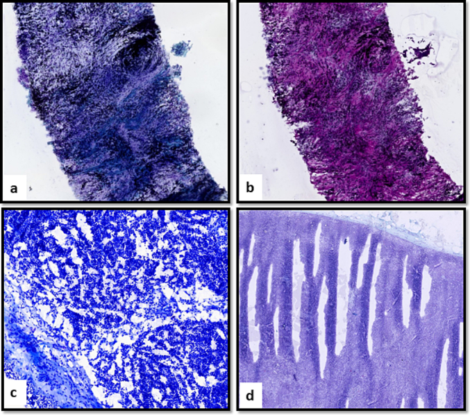
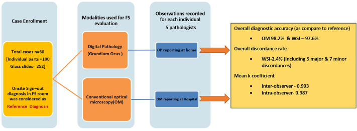
Cases accessioned beyond regular working hours (5 pm–10 pm) were reported simultaneously using optical microscopy (OM) and WSI. Validation of WSI for FS diagnosis from a remote site, i.e. home, was performed by 5 pathologists. Cases were scanned using a portable scanner (Grundium Ocus®40) and previewed on consumer-grade computer devices through a web-based browser (http://grundium.net). Clinical data and diagnostic reports were shared through a google spreadsheet. The diagnostic concordance, inter- and intra-observer agreement for FS diagnosis by WSI versus OM, and turnaround time (TAT), were recorded.
Results
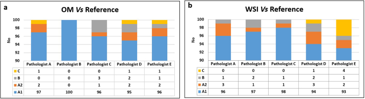
The overall diagnostic accuracy for OM and WSI (from home) was 98.2% (range 97%–100%) and 97.6% (range 95%–99%), respectively, when compared with the reference standard. Almost perfect inter-observer (k = 0.993) and intra-observer (k = 0.987) agreement for WSI was observed by 4 pathologists. Pathologists used consumer-grade laptops/desktops with an average screen size of 14.58 inches (range = 12.3–17.7 inches) and a network speed of 64 megabits per second (range: 10–90 Mbps). The mean diagnostic assessment time per case for OM and WSI was 1:48 min and 5:54 min, respectively. Mean TAT of 27.27 min per case was observed using WSI from home. Seamless connectivity was observed in approximately 75% of cases.
Conclusion
This study validates the role of WSI for remote FS diagnosis for its safe and efficient adoption in clinical use.
期刊介绍:
The Journal of Pathology Informatics (JPI) is an open access peer-reviewed journal dedicated to the advancement of pathology informatics. This is the official journal of the Association for Pathology Informatics (API). The journal aims to publish broadly about pathology informatics and freely disseminate all articles worldwide. This journal is of interest to pathologists, informaticians, academics, researchers, health IT specialists, information officers, IT staff, vendors, and anyone with an interest in informatics. We encourage submissions from anyone with an interest in the field of pathology informatics. We publish all types of papers related to pathology informatics including original research articles, technical notes, reviews, viewpoints, commentaries, editorials, symposia, meeting abstracts, book reviews, and correspondence to the editors. All submissions are subject to rigorous peer review by the well-regarded editorial board and by expert referees in appropriate specialties.


 分享
分享
 求助内容:
求助内容: 应助结果提醒方式:
 扫码关注我们
