{"title":"鸡鸭胃肠道的解剖、组织化学和免疫组织化学观察。","authors":"Ahmed M Abdellatif, Amany Farag, Elsayed Metwally","doi":"10.1186/s40850-022-00161-6","DOIUrl":null,"url":null,"abstract":"<p><strong>Background: </strong>Gallinula chloropus (Linnaeus, 1758) is a wild aquatic omnivorous bird characterized by a marked resistance to harsh environmental conditions and a worldwide distribution. In this study, anatomical, morphometrical, histochemical, and immunohistochemical techniques were employed to study the structure of the gastrointestinal tract of Gallinula chloropus.</p><p><strong>Results: </strong>The esophagus appeared tubular with no distinct crop. Both superficial (SPG) and deep (DPG) proventricular glands were present. The DPG filled about two-thirds of the total wall thickness. Histochemically, the mucosubstances revealed mixed alcian blue-PAS positive reactions. They were mainly localized in the acini of the esophageal glands and SPG, gastric surface epithelium, duct system of DPG, and intestinal goblet cells. The highest number of goblet cells per every 1 mm<sup>2</sup> of the intestinal mucosa was seen within the ileum and rectum, 2555 ± 468 and 2607 ± 653 respectively. Notably, glucagon immunoreactive (IR) cells were abundant in the mucosa of the small and large intestines and the proventriculus, while somatostatin IR cells were concentrated within the acini of the DPG. IR cells for the mitosis marker phospho-histone H3 (PHH3) were highest within the entire intestinal crypts and mucosa-associated lymphoid tissues (MALT). In contrast, cells IR for the apoptosis marker C.CASP3 were remarkable in epithelial cells at the tips of intestinal villi and in MALT, reflecting the dynamic nature of the latter mentioned structures.</p><p><strong>Conclusions: </strong>The findings of the present study advance our knowledge of the gross and microscopic anatomy of the gastrointestinal tract in wild birds and could help to enhance the productivity of Aves via improving gut health.</p>","PeriodicalId":48590,"journal":{"name":"BMC Zoology","volume":"7 1","pages":"61"},"PeriodicalIF":1.7000,"publicationDate":"2022-12-28","publicationTypes":"Journal Article","fieldsOfStudy":null,"isOpenAccess":false,"openAccessPdf":"https://www.ncbi.nlm.nih.gov/pmc/articles/PMC10127349/pdf/","citationCount":"0","resultStr":"{\"title\":\"Anatomical, histochemical, and immunohistochemical observations on the gastrointestinal tract of Gallinula chloropus (Aves: Rallidae).\",\"authors\":\"Ahmed M Abdellatif, Amany Farag, Elsayed Metwally\",\"doi\":\"10.1186/s40850-022-00161-6\",\"DOIUrl\":null,\"url\":null,\"abstract\":\"<p><strong>Background: </strong>Gallinula chloropus (Linnaeus, 1758) is a wild aquatic omnivorous bird characterized by a marked resistance to harsh environmental conditions and a worldwide distribution. In this study, anatomical, morphometrical, histochemical, and immunohistochemical techniques were employed to study the structure of the gastrointestinal tract of Gallinula chloropus.</p><p><strong>Results: </strong>The esophagus appeared tubular with no distinct crop. Both superficial (SPG) and deep (DPG) proventricular glands were present. The DPG filled about two-thirds of the total wall thickness. Histochemically, the mucosubstances revealed mixed alcian blue-PAS positive reactions. They were mainly localized in the acini of the esophageal glands and SPG, gastric surface epithelium, duct system of DPG, and intestinal goblet cells. The highest number of goblet cells per every 1 mm<sup>2</sup> of the intestinal mucosa was seen within the ileum and rectum, 2555 ± 468 and 2607 ± 653 respectively. Notably, glucagon immunoreactive (IR) cells were abundant in the mucosa of the small and large intestines and the proventriculus, while somatostatin IR cells were concentrated within the acini of the DPG. IR cells for the mitosis marker phospho-histone H3 (PHH3) were highest within the entire intestinal crypts and mucosa-associated lymphoid tissues (MALT). In contrast, cells IR for the apoptosis marker C.CASP3 were remarkable in epithelial cells at the tips of intestinal villi and in MALT, reflecting the dynamic nature of the latter mentioned structures.</p><p><strong>Conclusions: </strong>The findings of the present study advance our knowledge of the gross and microscopic anatomy of the gastrointestinal tract in wild birds and could help to enhance the productivity of Aves via improving gut health.</p>\",\"PeriodicalId\":48590,\"journal\":{\"name\":\"BMC Zoology\",\"volume\":\"7 1\",\"pages\":\"61\"},\"PeriodicalIF\":1.7000,\"publicationDate\":\"2022-12-28\",\"publicationTypes\":\"Journal Article\",\"fieldsOfStudy\":null,\"isOpenAccess\":false,\"openAccessPdf\":\"https://www.ncbi.nlm.nih.gov/pmc/articles/PMC10127349/pdf/\",\"citationCount\":\"0\",\"resultStr\":null,\"platform\":\"Semanticscholar\",\"paperid\":null,\"PeriodicalName\":\"BMC Zoology\",\"FirstCategoryId\":\"99\",\"ListUrlMain\":\"https://doi.org/10.1186/s40850-022-00161-6\",\"RegionNum\":3,\"RegionCategory\":\"生物学\",\"ArticlePicture\":[],\"TitleCN\":null,\"AbstractTextCN\":null,\"PMCID\":null,\"EPubDate\":\"\",\"PubModel\":\"\",\"JCR\":\"Q2\",\"JCRName\":\"ZOOLOGY\",\"Score\":null,\"Total\":0}","platform":"Semanticscholar","paperid":null,"PeriodicalName":"BMC Zoology","FirstCategoryId":"99","ListUrlMain":"https://doi.org/10.1186/s40850-022-00161-6","RegionNum":3,"RegionCategory":"生物学","ArticlePicture":[],"TitleCN":null,"AbstractTextCN":null,"PMCID":null,"EPubDate":"","PubModel":"","JCR":"Q2","JCRName":"ZOOLOGY","Score":null,"Total":0}
Anatomical, histochemical, and immunohistochemical observations on the gastrointestinal tract of Gallinula chloropus (Aves: Rallidae).
Background: Gallinula chloropus (Linnaeus, 1758) is a wild aquatic omnivorous bird characterized by a marked resistance to harsh environmental conditions and a worldwide distribution. In this study, anatomical, morphometrical, histochemical, and immunohistochemical techniques were employed to study the structure of the gastrointestinal tract of Gallinula chloropus.
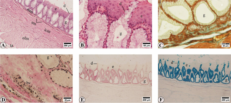
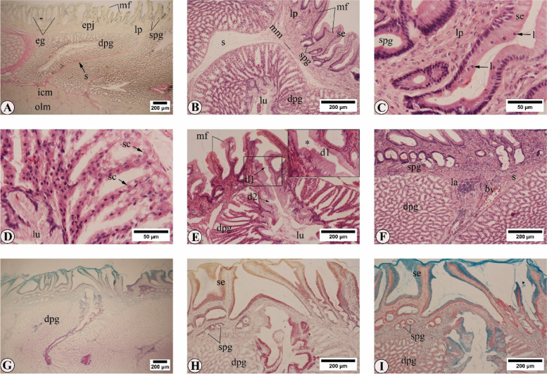
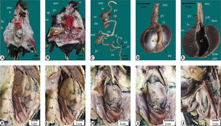
Results: The esophagus appeared tubular with no distinct crop. Both superficial (SPG) and deep (DPG) proventricular glands were present. The DPG filled about two-thirds of the total wall thickness. Histochemically, the mucosubstances revealed mixed alcian blue-PAS positive reactions. They were mainly localized in the acini of the esophageal glands and SPG, gastric surface epithelium, duct system of DPG, and intestinal goblet cells. The highest number of goblet cells per every 1 mm2 of the intestinal mucosa was seen within the ileum and rectum, 2555 ± 468 and 2607 ± 653 respectively. Notably, glucagon immunoreactive (IR) cells were abundant in the mucosa of the small and large intestines and the proventriculus, while somatostatin IR cells were concentrated within the acini of the DPG. IR cells for the mitosis marker phospho-histone H3 (PHH3) were highest within the entire intestinal crypts and mucosa-associated lymphoid tissues (MALT). In contrast, cells IR for the apoptosis marker C.CASP3 were remarkable in epithelial cells at the tips of intestinal villi and in MALT, reflecting the dynamic nature of the latter mentioned structures.
Conclusions: The findings of the present study advance our knowledge of the gross and microscopic anatomy of the gastrointestinal tract in wild birds and could help to enhance the productivity of Aves via improving gut health.
BMC ZoologyAgricultural and Biological Sciences-Animal Science and Zoology
CiteScore
2.30
自引率
6.20%
发文量
53
审稿时长
24 weeks
期刊介绍:
BMC Zoology is an open access, peer-reviewed journal that considers articles on all aspects of zoology, including physiology, mechanistic and functional studies, anatomy, life history, behavior, signalling and communication, cognition, parasitism, taxonomy and conservation.


 分享
分享
 求助内容:
求助内容: 应助结果提醒方式:
 扫码关注我们
