Ehsan Safai Zadeh, Amjad Alhyari, Johannes Kroenig, Christian Görg, Corinna Trenker, Christoph F. Dietrich, Hajo Findeisen
{"title":"b超和增强超声对肺炎的评价:一篇图片文章","authors":"Ehsan Safai Zadeh, Amjad Alhyari, Johannes Kroenig, Christian Görg, Corinna Trenker, Christoph F. Dietrich, Hajo Findeisen","doi":"10.1002/ajum.12332","DOIUrl":null,"url":null,"abstract":"<p>Due to their often peripheral pleural-based location, pneumonias can be visualised by B-mode ultrasound. Therefore, sonography can be used as an alternative imaging modality to chest X-ray in suspected cases of pneumonia. Depending on the clinical background of the patient, and various underlying pathological mechanisms, a heterogeneous pattern of pneumonia is seen in both B-mode lung ultrasound and contrast-enhanced ultrasound. Here, we describe the spectrum of sonographic manifestations of pneumonic/inflammatory consolidation on B-mode lung ultrasound and contrast-enhanced ultrasound.</p>","PeriodicalId":36517,"journal":{"name":"Australasian Journal of Ultrasound in Medicine","volume":"26 2","pages":"100-114"},"PeriodicalIF":0.0000,"publicationDate":"2023-02-15","publicationTypes":"Journal Article","fieldsOfStudy":null,"isOpenAccess":false,"openAccessPdf":"https://onlinelibrary.wiley.com/doi/epdf/10.1002/ajum.12332","citationCount":"2","resultStr":"{\"title\":\"B-mode ultrasound and contrast-enhanced ultrasound for evaluation of pneumonia: A pictorial essay\",\"authors\":\"Ehsan Safai Zadeh, Amjad Alhyari, Johannes Kroenig, Christian Görg, Corinna Trenker, Christoph F. Dietrich, Hajo Findeisen\",\"doi\":\"10.1002/ajum.12332\",\"DOIUrl\":null,\"url\":null,\"abstract\":\"<p>Due to their often peripheral pleural-based location, pneumonias can be visualised by B-mode ultrasound. Therefore, sonography can be used as an alternative imaging modality to chest X-ray in suspected cases of pneumonia. Depending on the clinical background of the patient, and various underlying pathological mechanisms, a heterogeneous pattern of pneumonia is seen in both B-mode lung ultrasound and contrast-enhanced ultrasound. Here, we describe the spectrum of sonographic manifestations of pneumonic/inflammatory consolidation on B-mode lung ultrasound and contrast-enhanced ultrasound.</p>\",\"PeriodicalId\":36517,\"journal\":{\"name\":\"Australasian Journal of Ultrasound in Medicine\",\"volume\":\"26 2\",\"pages\":\"100-114\"},\"PeriodicalIF\":0.0000,\"publicationDate\":\"2023-02-15\",\"publicationTypes\":\"Journal Article\",\"fieldsOfStudy\":null,\"isOpenAccess\":false,\"openAccessPdf\":\"https://onlinelibrary.wiley.com/doi/epdf/10.1002/ajum.12332\",\"citationCount\":\"2\",\"resultStr\":null,\"platform\":\"Semanticscholar\",\"paperid\":null,\"PeriodicalName\":\"Australasian Journal of Ultrasound in Medicine\",\"FirstCategoryId\":\"1085\",\"ListUrlMain\":\"https://onlinelibrary.wiley.com/doi/10.1002/ajum.12332\",\"RegionNum\":0,\"RegionCategory\":null,\"ArticlePicture\":[],\"TitleCN\":null,\"AbstractTextCN\":null,\"PMCID\":null,\"EPubDate\":\"\",\"PubModel\":\"\",\"JCR\":\"Q3\",\"JCRName\":\"Medicine\",\"Score\":null,\"Total\":0}","platform":"Semanticscholar","paperid":null,"PeriodicalName":"Australasian Journal of Ultrasound in Medicine","FirstCategoryId":"1085","ListUrlMain":"https://onlinelibrary.wiley.com/doi/10.1002/ajum.12332","RegionNum":0,"RegionCategory":null,"ArticlePicture":[],"TitleCN":null,"AbstractTextCN":null,"PMCID":null,"EPubDate":"","PubModel":"","JCR":"Q3","JCRName":"Medicine","Score":null,"Total":0}
B-mode ultrasound and contrast-enhanced ultrasound for evaluation of pneumonia: A pictorial essay
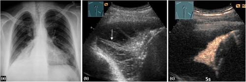
Due to their often peripheral pleural-based location, pneumonias can be visualised by B-mode ultrasound. Therefore, sonography can be used as an alternative imaging modality to chest X-ray in suspected cases of pneumonia. Depending on the clinical background of the patient, and various underlying pathological mechanisms, a heterogeneous pattern of pneumonia is seen in both B-mode lung ultrasound and contrast-enhanced ultrasound. Here, we describe the spectrum of sonographic manifestations of pneumonic/inflammatory consolidation on B-mode lung ultrasound and contrast-enhanced ultrasound.


 分享
分享
 求助内容:
求助内容: 应助结果提醒方式:
 扫码关注我们
