Luciano Augusto Cano Martins, Danieli Moura Brasil, Deborah Queiroz Freitas, Matheus L Oliveira
{"title":"一种利用锥束计算机断层扫描客观检测牙齿强直的新方法:实验室研究。","authors":"Luciano Augusto Cano Martins, Danieli Moura Brasil, Deborah Queiroz Freitas, Matheus L Oliveira","doi":"10.5624/isd.20220186","DOIUrl":null,"url":null,"abstract":"<p><strong>Purpose: </strong>The aim of this study was to objectively detect simulated tooth ankylosis using a novel method involving cone-beam computed tomography (CBCT).</p><p><strong>Materials and methods: </strong>Tooth ankylosis was simulated in single-rooted human permanent teeth, and CBCT scans were acquired at different current levels (5, 6.3, and 8 mA) and voxel sizes (0.08, 0.125, and 0.2). In axial reconstructions, a line of interest was perpendicularly placed over the periodontal ligament space of 21 ankylosed and 21 non-ankylosed regions, and the CBCT grey values of all voxels along the line of interest were plotted against their corresponding X-coordinates through a line graph to generate a profile. The image contrast was increased by 30% and 60% and the profile assessment was repeated. The internal area of the resulting parabolas was obtained from all images and compared between ankylosed and non-ankylosed regions under different contrast enhancement conditions, voxel sizes, and mA levels using multi-way analysis of variance with the Tukey <i>post hoc</i> test (α=0.05).</p><p><strong>Results: </strong>The internal area of the parabolas of all non-ankylosed regions was significantly higher than that of the ankylosed regions (<i>P</i><0.05). Contrast enhancement led to a significantly greater internal area of the parabolas of non-ankylosed regions (<i>P</i><0.05). Overall, voxel size and mA did not significantly influence the internal area of the parabolas (<i>P</i>>0.05).</p><p><strong>Conclusion: </strong>The proposed novel method revealed a relevant degree of applicability in the detection of simulated tooth ankylosis; increased image contrast led to greater detectability.</p>","PeriodicalId":51714,"journal":{"name":"Imaging Science in Dentistry","volume":"53 1","pages":"61-67"},"PeriodicalIF":2.1000,"publicationDate":"2023-03-01","publicationTypes":"Journal Article","fieldsOfStudy":null,"isOpenAccess":false,"openAccessPdf":"https://ftp.ncbi.nlm.nih.gov/pub/pmc/oa_pdf/d2/b4/isd-53-61.PMC10060758.pdf","citationCount":"0","resultStr":"{\"title\":\"A novel method of objectively detecting tooth ankylosis using cone-beam computed tomography: A laboratory study.\",\"authors\":\"Luciano Augusto Cano Martins, Danieli Moura Brasil, Deborah Queiroz Freitas, Matheus L Oliveira\",\"doi\":\"10.5624/isd.20220186\",\"DOIUrl\":null,\"url\":null,\"abstract\":\"<p><strong>Purpose: </strong>The aim of this study was to objectively detect simulated tooth ankylosis using a novel method involving cone-beam computed tomography (CBCT).</p><p><strong>Materials and methods: </strong>Tooth ankylosis was simulated in single-rooted human permanent teeth, and CBCT scans were acquired at different current levels (5, 6.3, and 8 mA) and voxel sizes (0.08, 0.125, and 0.2). In axial reconstructions, a line of interest was perpendicularly placed over the periodontal ligament space of 21 ankylosed and 21 non-ankylosed regions, and the CBCT grey values of all voxels along the line of interest were plotted against their corresponding X-coordinates through a line graph to generate a profile. The image contrast was increased by 30% and 60% and the profile assessment was repeated. The internal area of the resulting parabolas was obtained from all images and compared between ankylosed and non-ankylosed regions under different contrast enhancement conditions, voxel sizes, and mA levels using multi-way analysis of variance with the Tukey <i>post hoc</i> test (α=0.05).</p><p><strong>Results: </strong>The internal area of the parabolas of all non-ankylosed regions was significantly higher than that of the ankylosed regions (<i>P</i><0.05). Contrast enhancement led to a significantly greater internal area of the parabolas of non-ankylosed regions (<i>P</i><0.05). Overall, voxel size and mA did not significantly influence the internal area of the parabolas (<i>P</i>>0.05).</p><p><strong>Conclusion: </strong>The proposed novel method revealed a relevant degree of applicability in the detection of simulated tooth ankylosis; increased image contrast led to greater detectability.</p>\",\"PeriodicalId\":51714,\"journal\":{\"name\":\"Imaging Science in Dentistry\",\"volume\":\"53 1\",\"pages\":\"61-67\"},\"PeriodicalIF\":2.1000,\"publicationDate\":\"2023-03-01\",\"publicationTypes\":\"Journal Article\",\"fieldsOfStudy\":null,\"isOpenAccess\":false,\"openAccessPdf\":\"https://ftp.ncbi.nlm.nih.gov/pub/pmc/oa_pdf/d2/b4/isd-53-61.PMC10060758.pdf\",\"citationCount\":\"0\",\"resultStr\":null,\"platform\":\"Semanticscholar\",\"paperid\":null,\"PeriodicalName\":\"Imaging Science in Dentistry\",\"FirstCategoryId\":\"1085\",\"ListUrlMain\":\"https://doi.org/10.5624/isd.20220186\",\"RegionNum\":0,\"RegionCategory\":null,\"ArticlePicture\":[],\"TitleCN\":null,\"AbstractTextCN\":null,\"PMCID\":null,\"EPubDate\":\"\",\"PubModel\":\"\",\"JCR\":\"Q3\",\"JCRName\":\"DENTISTRY, ORAL SURGERY & MEDICINE\",\"Score\":null,\"Total\":0}","platform":"Semanticscholar","paperid":null,"PeriodicalName":"Imaging Science in Dentistry","FirstCategoryId":"1085","ListUrlMain":"https://doi.org/10.5624/isd.20220186","RegionNum":0,"RegionCategory":null,"ArticlePicture":[],"TitleCN":null,"AbstractTextCN":null,"PMCID":null,"EPubDate":"","PubModel":"","JCR":"Q3","JCRName":"DENTISTRY, ORAL SURGERY & MEDICINE","Score":null,"Total":0}
A novel method of objectively detecting tooth ankylosis using cone-beam computed tomography: A laboratory study.
Purpose: The aim of this study was to objectively detect simulated tooth ankylosis using a novel method involving cone-beam computed tomography (CBCT).
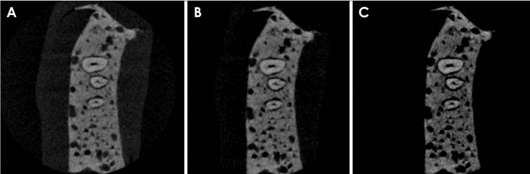
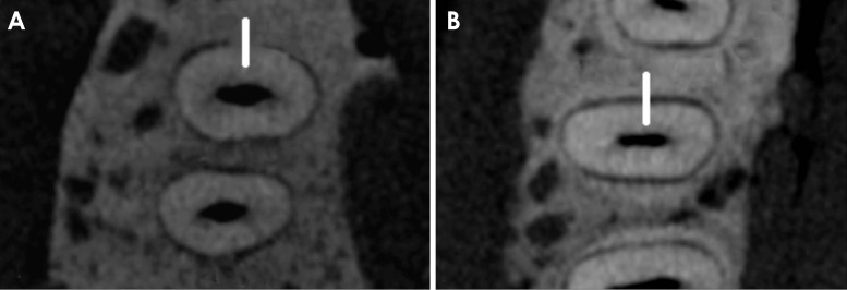
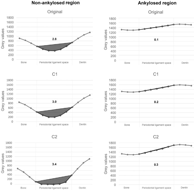
Materials and methods: Tooth ankylosis was simulated in single-rooted human permanent teeth, and CBCT scans were acquired at different current levels (5, 6.3, and 8 mA) and voxel sizes (0.08, 0.125, and 0.2). In axial reconstructions, a line of interest was perpendicularly placed over the periodontal ligament space of 21 ankylosed and 21 non-ankylosed regions, and the CBCT grey values of all voxels along the line of interest were plotted against their corresponding X-coordinates through a line graph to generate a profile. The image contrast was increased by 30% and 60% and the profile assessment was repeated. The internal area of the resulting parabolas was obtained from all images and compared between ankylosed and non-ankylosed regions under different contrast enhancement conditions, voxel sizes, and mA levels using multi-way analysis of variance with the Tukey post hoc test (α=0.05).
Results: The internal area of the parabolas of all non-ankylosed regions was significantly higher than that of the ankylosed regions (P<0.05). Contrast enhancement led to a significantly greater internal area of the parabolas of non-ankylosed regions (P<0.05). Overall, voxel size and mA did not significantly influence the internal area of the parabolas (P>0.05).
Conclusion: The proposed novel method revealed a relevant degree of applicability in the detection of simulated tooth ankylosis; increased image contrast led to greater detectability.


 分享
分享
 求助内容:
求助内容: 应助结果提醒方式:
 扫码关注我们
