Hyung In Choi, You Me Kim, Junwon Min, Yong Moon Lee, Hee Jeong Kim
{"title":"原发性乳腺恶性黑色素瘤表现为乳腺脓肿:1例报告。","authors":"Hyung In Choi, You Me Kim, Junwon Min, Yong Moon Lee, Hee Jeong Kim","doi":"10.3348/jksr.2022.0014","DOIUrl":null,"url":null,"abstract":"<p><p>Primary malignant melanoma in breast parenchyma (PMB) is an extremely rare disease, and the most common presentation is a palpable breast lump. To the best of our knowledge, a case of PMB presenting as a breast abscess has not been reported in English literatures. We present a case of PMB that manifested as a recurrent breast abscess in a 71-year-old woman. On MRI, an enhancing solid mass with a cystic or necrotic portion was revealed with some high signal intensities on precontrast-enhanced T1-weighted images and a dark rim on T2-weighed images. The MRI features played a pivotal role in identifying the underlying malignant condition and making an accurate diagnosis of this rare case of PMB with unusual clinical presentation.</p>","PeriodicalId":17455,"journal":{"name":"Journal of the Korean Society of Radiology","volume":"84 3","pages":"763-769"},"PeriodicalIF":0.0000,"publicationDate":"2023-05-01","publicationTypes":"Journal Article","fieldsOfStudy":null,"isOpenAccess":false,"openAccessPdf":"https://ftp.ncbi.nlm.nih.gov/pub/pmc/oa_pdf/05/0b/jksr-84-763.PMC10265238.pdf","citationCount":"0","resultStr":"{\"title\":\"Primary Malignant Melanoma of the Breast Presenting as a Breast Abscess: A Case Report.\",\"authors\":\"Hyung In Choi, You Me Kim, Junwon Min, Yong Moon Lee, Hee Jeong Kim\",\"doi\":\"10.3348/jksr.2022.0014\",\"DOIUrl\":null,\"url\":null,\"abstract\":\"<p><p>Primary malignant melanoma in breast parenchyma (PMB) is an extremely rare disease, and the most common presentation is a palpable breast lump. To the best of our knowledge, a case of PMB presenting as a breast abscess has not been reported in English literatures. We present a case of PMB that manifested as a recurrent breast abscess in a 71-year-old woman. On MRI, an enhancing solid mass with a cystic or necrotic portion was revealed with some high signal intensities on precontrast-enhanced T1-weighted images and a dark rim on T2-weighed images. The MRI features played a pivotal role in identifying the underlying malignant condition and making an accurate diagnosis of this rare case of PMB with unusual clinical presentation.</p>\",\"PeriodicalId\":17455,\"journal\":{\"name\":\"Journal of the Korean Society of Radiology\",\"volume\":\"84 3\",\"pages\":\"763-769\"},\"PeriodicalIF\":0.0000,\"publicationDate\":\"2023-05-01\",\"publicationTypes\":\"Journal Article\",\"fieldsOfStudy\":null,\"isOpenAccess\":false,\"openAccessPdf\":\"https://ftp.ncbi.nlm.nih.gov/pub/pmc/oa_pdf/05/0b/jksr-84-763.PMC10265238.pdf\",\"citationCount\":\"0\",\"resultStr\":null,\"platform\":\"Semanticscholar\",\"paperid\":null,\"PeriodicalName\":\"Journal of the Korean Society of Radiology\",\"FirstCategoryId\":\"1085\",\"ListUrlMain\":\"https://doi.org/10.3348/jksr.2022.0014\",\"RegionNum\":0,\"RegionCategory\":null,\"ArticlePicture\":[],\"TitleCN\":null,\"AbstractTextCN\":null,\"PMCID\":null,\"EPubDate\":\"\",\"PubModel\":\"\",\"JCR\":\"Q4\",\"JCRName\":\"Medicine\",\"Score\":null,\"Total\":0}","platform":"Semanticscholar","paperid":null,"PeriodicalName":"Journal of the Korean Society of Radiology","FirstCategoryId":"1085","ListUrlMain":"https://doi.org/10.3348/jksr.2022.0014","RegionNum":0,"RegionCategory":null,"ArticlePicture":[],"TitleCN":null,"AbstractTextCN":null,"PMCID":null,"EPubDate":"","PubModel":"","JCR":"Q4","JCRName":"Medicine","Score":null,"Total":0}
Primary Malignant Melanoma of the Breast Presenting as a Breast Abscess: A Case Report.
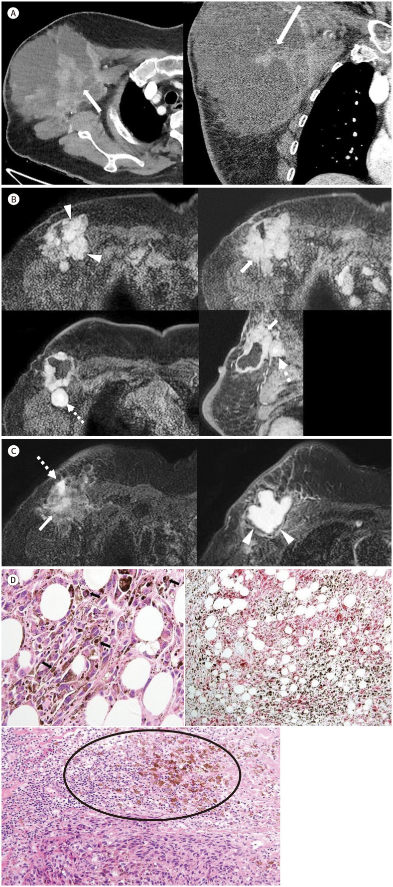
Primary malignant melanoma in breast parenchyma (PMB) is an extremely rare disease, and the most common presentation is a palpable breast lump. To the best of our knowledge, a case of PMB presenting as a breast abscess has not been reported in English literatures. We present a case of PMB that manifested as a recurrent breast abscess in a 71-year-old woman. On MRI, an enhancing solid mass with a cystic or necrotic portion was revealed with some high signal intensities on precontrast-enhanced T1-weighted images and a dark rim on T2-weighed images. The MRI features played a pivotal role in identifying the underlying malignant condition and making an accurate diagnosis of this rare case of PMB with unusual clinical presentation.


 分享
分享
 求助内容:
求助内容: 应助结果提醒方式:
 扫码关注我们
