{"title":"[利用VX2细胞建立兔脑肿瘤模型及其在神经放射学研究中的MRI验证]。","authors":"Yong-Woo Kim, Seon Hee Choi, Hak Jin Kim","doi":"10.3348/jksr.2022.0078","DOIUrl":null,"url":null,"abstract":"<p><strong>Purpose: </strong>To evaluate the development, location, and volume of a VX2 carcinoma using four inoculation methods in a rabbit brain.</p><p><strong>Materials and methods: </strong>Inoculation of a VX2 cell suspension was performed 1) on the appointed day, 2) seven days after storing a VX2 carcinoma in a freezer or 3) seven days after storing a VX2 carcinoma in a deep freezer after sacrificing the donor rabbits. 4) Without sacrificing the rabbits, the VX2 cell suspension was obtained using a gun biopsy, inoculation was performed on the appointed day. MR imaging was performed 10 days after inoculation. Brain tissues were obtained the day after. The development, location, and volume of the tumor were evaluated.</p><p><strong>Results: </strong>Seventeen of the 18 rabbits inoculated on the appointed day developed tumors (average tumor volume, 106.32 mm<sup>3</sup>). One of five inoculated seven days after storing the VX2 tumor in the freezer, and three of five inoculated seven days after storing the VX2 tumor in the deep freezer developed tumors. Inoculation with a VX2 cell suspension obtained with a gun biopsy from five rabbits revealed development of tumors in only two rabbits. The tumors mostly developed in the superficial cortex.</p><p><strong>Conclusion: </strong>TVX2 rabbit brain tumor model is easy to develop and revealed variable reproducibility. This model can be applicable in radiologic imaging, treatment planning, interventional treatment and drug delivery research. VX2 cell can be successfully innoculated into the brain using variable methods under researcher's variable conditions.</p>","PeriodicalId":17455,"journal":{"name":"Journal of the Korean Society of Radiology","volume":"84 2","pages":"441-453"},"PeriodicalIF":0.0000,"publicationDate":"2023-03-01","publicationTypes":"Journal Article","fieldsOfStudy":null,"isOpenAccess":false,"openAccessPdf":"https://ftp.ncbi.nlm.nih.gov/pub/pmc/oa_pdf/ed/0c/jksr-84-441.PMC10083627.pdf","citationCount":"0","resultStr":"{\"title\":\"[Development of Rabbit Brain Tumor Model Using VX2 Cells and Verification with the MRI in Neuroradiologic Research].\",\"authors\":\"Yong-Woo Kim, Seon Hee Choi, Hak Jin Kim\",\"doi\":\"10.3348/jksr.2022.0078\",\"DOIUrl\":null,\"url\":null,\"abstract\":\"<p><strong>Purpose: </strong>To evaluate the development, location, and volume of a VX2 carcinoma using four inoculation methods in a rabbit brain.</p><p><strong>Materials and methods: </strong>Inoculation of a VX2 cell suspension was performed 1) on the appointed day, 2) seven days after storing a VX2 carcinoma in a freezer or 3) seven days after storing a VX2 carcinoma in a deep freezer after sacrificing the donor rabbits. 4) Without sacrificing the rabbits, the VX2 cell suspension was obtained using a gun biopsy, inoculation was performed on the appointed day. MR imaging was performed 10 days after inoculation. Brain tissues were obtained the day after. The development, location, and volume of the tumor were evaluated.</p><p><strong>Results: </strong>Seventeen of the 18 rabbits inoculated on the appointed day developed tumors (average tumor volume, 106.32 mm<sup>3</sup>). One of five inoculated seven days after storing the VX2 tumor in the freezer, and three of five inoculated seven days after storing the VX2 tumor in the deep freezer developed tumors. Inoculation with a VX2 cell suspension obtained with a gun biopsy from five rabbits revealed development of tumors in only two rabbits. The tumors mostly developed in the superficial cortex.</p><p><strong>Conclusion: </strong>TVX2 rabbit brain tumor model is easy to develop and revealed variable reproducibility. This model can be applicable in radiologic imaging, treatment planning, interventional treatment and drug delivery research. VX2 cell can be successfully innoculated into the brain using variable methods under researcher's variable conditions.</p>\",\"PeriodicalId\":17455,\"journal\":{\"name\":\"Journal of the Korean Society of Radiology\",\"volume\":\"84 2\",\"pages\":\"441-453\"},\"PeriodicalIF\":0.0000,\"publicationDate\":\"2023-03-01\",\"publicationTypes\":\"Journal Article\",\"fieldsOfStudy\":null,\"isOpenAccess\":false,\"openAccessPdf\":\"https://ftp.ncbi.nlm.nih.gov/pub/pmc/oa_pdf/ed/0c/jksr-84-441.PMC10083627.pdf\",\"citationCount\":\"0\",\"resultStr\":null,\"platform\":\"Semanticscholar\",\"paperid\":null,\"PeriodicalName\":\"Journal of the Korean Society of Radiology\",\"FirstCategoryId\":\"1085\",\"ListUrlMain\":\"https://doi.org/10.3348/jksr.2022.0078\",\"RegionNum\":0,\"RegionCategory\":null,\"ArticlePicture\":[],\"TitleCN\":null,\"AbstractTextCN\":null,\"PMCID\":null,\"EPubDate\":\"\",\"PubModel\":\"\",\"JCR\":\"Q4\",\"JCRName\":\"Medicine\",\"Score\":null,\"Total\":0}","platform":"Semanticscholar","paperid":null,"PeriodicalName":"Journal of the Korean Society of Radiology","FirstCategoryId":"1085","ListUrlMain":"https://doi.org/10.3348/jksr.2022.0078","RegionNum":0,"RegionCategory":null,"ArticlePicture":[],"TitleCN":null,"AbstractTextCN":null,"PMCID":null,"EPubDate":"","PubModel":"","JCR":"Q4","JCRName":"Medicine","Score":null,"Total":0}
[Development of Rabbit Brain Tumor Model Using VX2 Cells and Verification with the MRI in Neuroradiologic Research].
Purpose: To evaluate the development, location, and volume of a VX2 carcinoma using four inoculation methods in a rabbit brain.
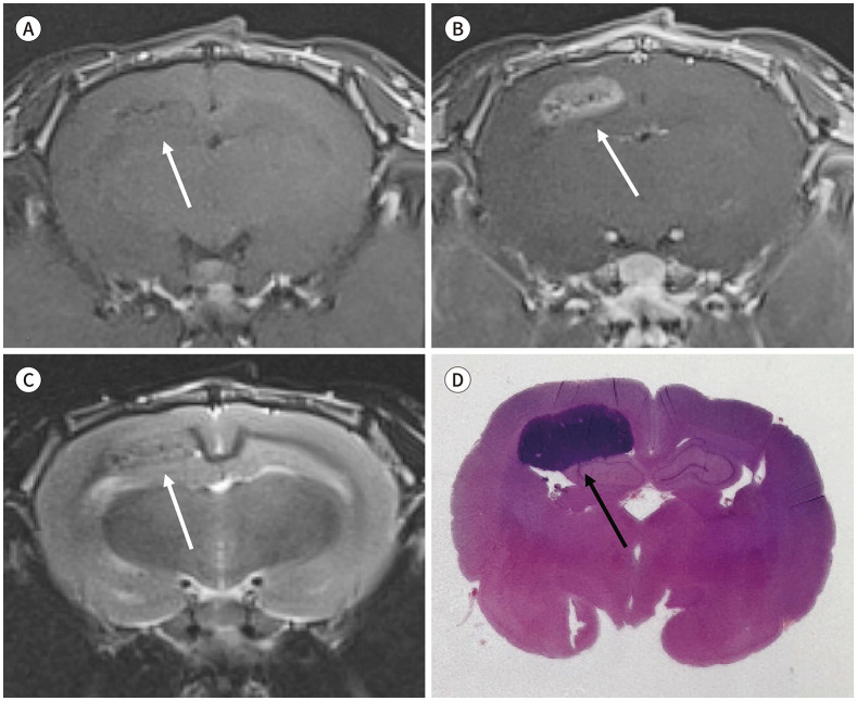
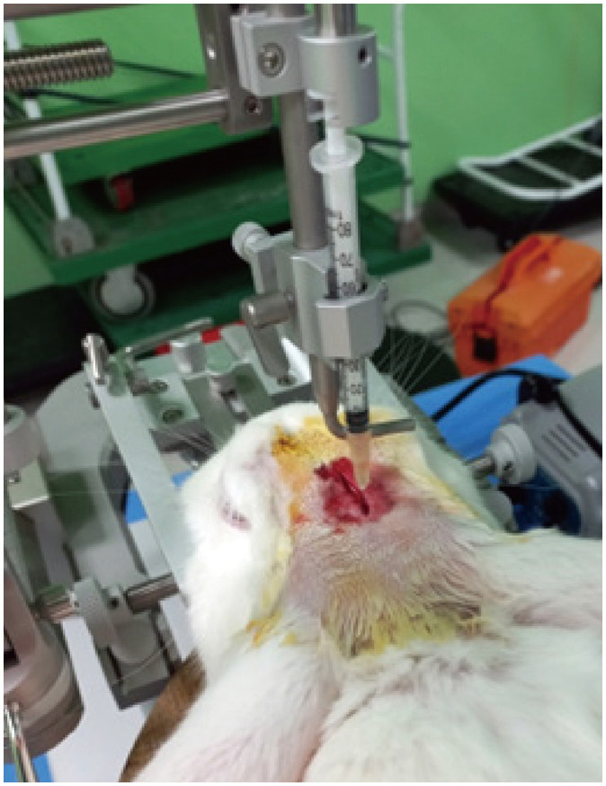
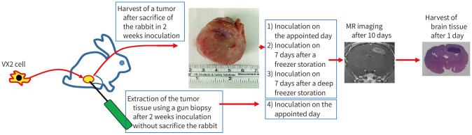
Materials and methods: Inoculation of a VX2 cell suspension was performed 1) on the appointed day, 2) seven days after storing a VX2 carcinoma in a freezer or 3) seven days after storing a VX2 carcinoma in a deep freezer after sacrificing the donor rabbits. 4) Without sacrificing the rabbits, the VX2 cell suspension was obtained using a gun biopsy, inoculation was performed on the appointed day. MR imaging was performed 10 days after inoculation. Brain tissues were obtained the day after. The development, location, and volume of the tumor were evaluated.
Results: Seventeen of the 18 rabbits inoculated on the appointed day developed tumors (average tumor volume, 106.32 mm3). One of five inoculated seven days after storing the VX2 tumor in the freezer, and three of five inoculated seven days after storing the VX2 tumor in the deep freezer developed tumors. Inoculation with a VX2 cell suspension obtained with a gun biopsy from five rabbits revealed development of tumors in only two rabbits. The tumors mostly developed in the superficial cortex.
Conclusion: TVX2 rabbit brain tumor model is easy to develop and revealed variable reproducibility. This model can be applicable in radiologic imaging, treatment planning, interventional treatment and drug delivery research. VX2 cell can be successfully innoculated into the brain using variable methods under researcher's variable conditions.


 分享
分享
 求助内容:
求助内容: 应助结果提醒方式:
 扫码关注我们
