Mohammad Wehbi, Evelyne Harkemanne, Lionel Mignion, Nicolas Joudiou, Isabelle Tromme, Jean-François Baurain, Bernard Gallez
{"title":"通过电子顺磁共振 (EPR) 光谱和黑色素成像确定临床皮肤黑色素瘤的特征。","authors":"Mohammad Wehbi, Evelyne Harkemanne, Lionel Mignion, Nicolas Joudiou, Isabelle Tromme, Jean-François Baurain, Bernard Gallez","doi":"10.1007/s11307-023-01836-3","DOIUrl":null,"url":null,"abstract":"<p><p>The incidence of melanoma is continuously increasing over time. Melanoma is the most aggressive skin cancer, significantly reducing quality of life and survival rates of patients at advanced stages. Therefore, early diagnosis remains the key to change the prognosis of patients with melanoma. In this context, advanced technologies are under evaluation to increase the accuracy of the diagnostic, to better characterize the lesions and visualize their possible invasiveness in the epidermis. Among the innovative methods, because melanin is paramagnetic, clinical low frequency electron paramagnetic resonance (EPR) that characterizes the melanin content in the lesion has the potential to be an adjunct diagnostic method of melanoma. In this review, we first summarize the challenges faced by dermatologists and oncologists in melanoma diagnostic and management. We also provide a historical perspective on melanin detection with a focus on EPR spectroscopy/imaging of melanomas. We describe key elements that allow EPR to move from in vitro studies to in vivo and finally to patients for melanoma studies. Finally, we provide a critical view on challenges to meet to make EPR operational in the clinic to characterize pigmented lesions.</p>","PeriodicalId":18760,"journal":{"name":"Molecular Imaging and Biology","volume":" ","pages":"382-390"},"PeriodicalIF":2.5000,"publicationDate":"2024-06-01","publicationTypes":"Journal Article","fieldsOfStudy":null,"isOpenAccess":false,"openAccessPdf":"https://www.ncbi.nlm.nih.gov/pmc/articles/PMC11211150/pdf/","citationCount":"0","resultStr":"{\"title\":\"Towards Characterization of Skin Melanoma in the Clinic by Electron Paramagnetic Resonance (EPR) Spectroscopy and Imaging of Melanin.\",\"authors\":\"Mohammad Wehbi, Evelyne Harkemanne, Lionel Mignion, Nicolas Joudiou, Isabelle Tromme, Jean-François Baurain, Bernard Gallez\",\"doi\":\"10.1007/s11307-023-01836-3\",\"DOIUrl\":null,\"url\":null,\"abstract\":\"<p><p>The incidence of melanoma is continuously increasing over time. Melanoma is the most aggressive skin cancer, significantly reducing quality of life and survival rates of patients at advanced stages. Therefore, early diagnosis remains the key to change the prognosis of patients with melanoma. In this context, advanced technologies are under evaluation to increase the accuracy of the diagnostic, to better characterize the lesions and visualize their possible invasiveness in the epidermis. Among the innovative methods, because melanin is paramagnetic, clinical low frequency electron paramagnetic resonance (EPR) that characterizes the melanin content in the lesion has the potential to be an adjunct diagnostic method of melanoma. In this review, we first summarize the challenges faced by dermatologists and oncologists in melanoma diagnostic and management. We also provide a historical perspective on melanin detection with a focus on EPR spectroscopy/imaging of melanomas. We describe key elements that allow EPR to move from in vitro studies to in vivo and finally to patients for melanoma studies. Finally, we provide a critical view on challenges to meet to make EPR operational in the clinic to characterize pigmented lesions.</p>\",\"PeriodicalId\":18760,\"journal\":{\"name\":\"Molecular Imaging and Biology\",\"volume\":\" \",\"pages\":\"382-390\"},\"PeriodicalIF\":2.5000,\"publicationDate\":\"2024-06-01\",\"publicationTypes\":\"Journal Article\",\"fieldsOfStudy\":null,\"isOpenAccess\":false,\"openAccessPdf\":\"https://www.ncbi.nlm.nih.gov/pmc/articles/PMC11211150/pdf/\",\"citationCount\":\"0\",\"resultStr\":null,\"platform\":\"Semanticscholar\",\"paperid\":null,\"PeriodicalName\":\"Molecular Imaging and Biology\",\"FirstCategoryId\":\"3\",\"ListUrlMain\":\"https://doi.org/10.1007/s11307-023-01836-3\",\"RegionNum\":4,\"RegionCategory\":\"医学\",\"ArticlePicture\":[],\"TitleCN\":null,\"AbstractTextCN\":null,\"PMCID\":null,\"EPubDate\":\"2023/6/30 0:00:00\",\"PubModel\":\"Epub\",\"JCR\":\"Q2\",\"JCRName\":\"RADIOLOGY, NUCLEAR MEDICINE & MEDICAL IMAGING\",\"Score\":null,\"Total\":0}","platform":"Semanticscholar","paperid":null,"PeriodicalName":"Molecular Imaging and Biology","FirstCategoryId":"3","ListUrlMain":"https://doi.org/10.1007/s11307-023-01836-3","RegionNum":4,"RegionCategory":"医学","ArticlePicture":[],"TitleCN":null,"AbstractTextCN":null,"PMCID":null,"EPubDate":"2023/6/30 0:00:00","PubModel":"Epub","JCR":"Q2","JCRName":"RADIOLOGY, NUCLEAR MEDICINE & MEDICAL IMAGING","Score":null,"Total":0}
Towards Characterization of Skin Melanoma in the Clinic by Electron Paramagnetic Resonance (EPR) Spectroscopy and Imaging of Melanin.
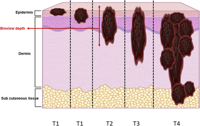
The incidence of melanoma is continuously increasing over time. Melanoma is the most aggressive skin cancer, significantly reducing quality of life and survival rates of patients at advanced stages. Therefore, early diagnosis remains the key to change the prognosis of patients with melanoma. In this context, advanced technologies are under evaluation to increase the accuracy of the diagnostic, to better characterize the lesions and visualize their possible invasiveness in the epidermis. Among the innovative methods, because melanin is paramagnetic, clinical low frequency electron paramagnetic resonance (EPR) that characterizes the melanin content in the lesion has the potential to be an adjunct diagnostic method of melanoma. In this review, we first summarize the challenges faced by dermatologists and oncologists in melanoma diagnostic and management. We also provide a historical perspective on melanin detection with a focus on EPR spectroscopy/imaging of melanomas. We describe key elements that allow EPR to move from in vitro studies to in vivo and finally to patients for melanoma studies. Finally, we provide a critical view on challenges to meet to make EPR operational in the clinic to characterize pigmented lesions.
期刊介绍:
Molecular Imaging and Biology (MIB) invites original contributions (research articles, review articles, commentaries, etc.) on the utilization of molecular imaging (i.e., nuclear imaging, optical imaging, autoradiography and pathology, MRI, MPI, ultrasound imaging, radiomics/genomics etc.) to investigate questions related to biology and health. The objective of MIB is to provide a forum to the discovery of molecular mechanisms of disease through the use of imaging techniques. We aim to investigate the biological nature of disease in patients and establish new molecular imaging diagnostic and therapy procedures.
Some areas that are covered are:
Preclinical and clinical imaging of macromolecular targets (e.g., genes, receptors, enzymes) involved in significant biological processes.
The design, characterization, and study of new molecular imaging probes and contrast agents for the functional interrogation of macromolecular targets.
Development and evaluation of imaging systems including instrumentation, image reconstruction algorithms, image analysis, and display.
Development of molecular assay approaches leading to quantification of the biological information obtained in molecular imaging.
Study of in vivo animal models of disease for the development of new molecular diagnostics and therapeutics.
Extension of in vitro and in vivo discoveries using disease models, into well designed clinical research investigations.
Clinical molecular imaging involving clinical investigations, clinical trials and medical management or cost-effectiveness studies.


 分享
分享

 求助内容:
求助内容: 应助结果提醒方式:
 扫码关注我们
