Barbara Knäusl , Peter Kuess , Markus Stock , Dietmar Georg , Piero Fossati , Petra Georg , Lukas Zimmermann
{"title":"在自适应碳离子处理工作流程中使用合成计算机断层扫描的可能性和挑战","authors":"Barbara Knäusl , Peter Kuess , Markus Stock , Dietmar Georg , Piero Fossati , Petra Georg , Lukas Zimmermann","doi":"10.1016/j.zemedi.2022.05.003","DOIUrl":null,"url":null,"abstract":"<div><h3>Background and purpose</h3><p>Anatomical surveillance during ion-beam therapy is the basis for an effective tumor treatment and optimal organ at risk (OAR) sparing. Synthetic computed tomography (sCT) based on magnetic resonance imaging (MRI) can replace the X-ray based planning CT (X-rayCT) in photon radiotherapy and improve the workflow efficiency without additional imaging dose. The extension to carbon-ion radiotherapy is highly challenging; complex patient positioning, unique anatomical situations, distinct horizontal and vertical beam incidence directions, and limited training data are only few problems. This study gives insight into the possibilities and challenges of using sCTs in carbon-ion therapy.</p></div><div><h3>Materials and methods</h3><p>For head and neck patients immobilised with thermoplastic masks 30 clinically applied actively scanned carbon-ion treatment plans on 15 CTs comprising 60 beams were analyzed. Those treatment plans were re-calculated on MRI based sCTs which were created employing a 3D U-Net. Dose differences and carbon-ion spot displacements between sCT and X-rayCT were evaluated on a patient specific basis.</p></div><div><h3>Results</h3><p>Spot displacement analysis showed a peak displacement by 0.2 cm caused by the immobilisation mask not measurable with the MRI. 95.7% of all spot displacements were located within 1 cm. For the clinical target volume (CTV) the median <span><math><mrow><msub><mrow><mi>D</mi></mrow><mrow><mn>50</mn><mo>%</mo></mrow></msub></mrow></math></span> agreed within −0.2% (−1.3 to 1.4%), while the median <span><math><mrow><msub><mrow><mi>D</mi></mrow><mrow><mn>0.01</mn><mspace></mspace><mi>cc</mi></mrow></msub></mrow></math></span> differed up to 4.2% (−1.3 to 25.3%) comparing the dose distribution on the X-rayCT and the sCT. OAR deviations depended strongly on the position and the dose gradient. For three patients no deterioration of the OAR parameters was observed. Other patients showed large deteriorations, <em>e.g.</em> for one patient <span><math><mrow><msub><mrow><mi>D</mi></mrow><mrow><mn>2</mn><mo>%</mo></mrow></msub></mrow></math></span> of the chiasm differed by 28.1%.</p></div><div><h3>Conclusion</h3><p>The usage of sCTs opens several new questions, concluding that we are not ready yet for an MR-only workflow in carbon-ion therapy, as envisaged in photon therapy. Although omitting the X-rayCT seems unfavourable in the case of carbon-ion therapy, an sCT could be advantageous for monitoring, re-planning, and adaptation.</p></div>","PeriodicalId":54397,"journal":{"name":"Zeitschrift fur Medizinische Physik","volume":"33 2","pages":"Pages 146-154"},"PeriodicalIF":4.2000,"publicationDate":"2023-05-01","publicationTypes":"Journal Article","fieldsOfStudy":null,"isOpenAccess":false,"openAccessPdf":"https://ftp.ncbi.nlm.nih.gov/pub/pmc/oa_pdf/8c/51/main.PMC10311249.pdf","citationCount":"2","resultStr":"{\"title\":\"Possibilities and challenges when using synthetic computed tomography in an adaptive carbon-ion treatment workflow\",\"authors\":\"Barbara Knäusl , Peter Kuess , Markus Stock , Dietmar Georg , Piero Fossati , Petra Georg , Lukas Zimmermann\",\"doi\":\"10.1016/j.zemedi.2022.05.003\",\"DOIUrl\":null,\"url\":null,\"abstract\":\"<div><h3>Background and purpose</h3><p>Anatomical surveillance during ion-beam therapy is the basis for an effective tumor treatment and optimal organ at risk (OAR) sparing. Synthetic computed tomography (sCT) based on magnetic resonance imaging (MRI) can replace the X-ray based planning CT (X-rayCT) in photon radiotherapy and improve the workflow efficiency without additional imaging dose. The extension to carbon-ion radiotherapy is highly challenging; complex patient positioning, unique anatomical situations, distinct horizontal and vertical beam incidence directions, and limited training data are only few problems. This study gives insight into the possibilities and challenges of using sCTs in carbon-ion therapy.</p></div><div><h3>Materials and methods</h3><p>For head and neck patients immobilised with thermoplastic masks 30 clinically applied actively scanned carbon-ion treatment plans on 15 CTs comprising 60 beams were analyzed. Those treatment plans were re-calculated on MRI based sCTs which were created employing a 3D U-Net. Dose differences and carbon-ion spot displacements between sCT and X-rayCT were evaluated on a patient specific basis.</p></div><div><h3>Results</h3><p>Spot displacement analysis showed a peak displacement by 0.2 cm caused by the immobilisation mask not measurable with the MRI. 95.7% of all spot displacements were located within 1 cm. For the clinical target volume (CTV) the median <span><math><mrow><msub><mrow><mi>D</mi></mrow><mrow><mn>50</mn><mo>%</mo></mrow></msub></mrow></math></span> agreed within −0.2% (−1.3 to 1.4%), while the median <span><math><mrow><msub><mrow><mi>D</mi></mrow><mrow><mn>0.01</mn><mspace></mspace><mi>cc</mi></mrow></msub></mrow></math></span> differed up to 4.2% (−1.3 to 25.3%) comparing the dose distribution on the X-rayCT and the sCT. OAR deviations depended strongly on the position and the dose gradient. For three patients no deterioration of the OAR parameters was observed. Other patients showed large deteriorations, <em>e.g.</em> for one patient <span><math><mrow><msub><mrow><mi>D</mi></mrow><mrow><mn>2</mn><mo>%</mo></mrow></msub></mrow></math></span> of the chiasm differed by 28.1%.</p></div><div><h3>Conclusion</h3><p>The usage of sCTs opens several new questions, concluding that we are not ready yet for an MR-only workflow in carbon-ion therapy, as envisaged in photon therapy. Although omitting the X-rayCT seems unfavourable in the case of carbon-ion therapy, an sCT could be advantageous for monitoring, re-planning, and adaptation.</p></div>\",\"PeriodicalId\":54397,\"journal\":{\"name\":\"Zeitschrift fur Medizinische Physik\",\"volume\":\"33 2\",\"pages\":\"Pages 146-154\"},\"PeriodicalIF\":4.2000,\"publicationDate\":\"2023-05-01\",\"publicationTypes\":\"Journal Article\",\"fieldsOfStudy\":null,\"isOpenAccess\":false,\"openAccessPdf\":\"https://ftp.ncbi.nlm.nih.gov/pub/pmc/oa_pdf/8c/51/main.PMC10311249.pdf\",\"citationCount\":\"2\",\"resultStr\":null,\"platform\":\"Semanticscholar\",\"paperid\":null,\"PeriodicalName\":\"Zeitschrift fur Medizinische Physik\",\"FirstCategoryId\":\"3\",\"ListUrlMain\":\"https://www.sciencedirect.com/science/article/pii/S0939388922000642\",\"RegionNum\":4,\"RegionCategory\":\"医学\",\"ArticlePicture\":[],\"TitleCN\":null,\"AbstractTextCN\":null,\"PMCID\":null,\"EPubDate\":\"\",\"PubModel\":\"\",\"JCR\":\"Q2\",\"JCRName\":\"RADIOLOGY, NUCLEAR MEDICINE & MEDICAL IMAGING\",\"Score\":null,\"Total\":0}","platform":"Semanticscholar","paperid":null,"PeriodicalName":"Zeitschrift fur Medizinische Physik","FirstCategoryId":"3","ListUrlMain":"https://www.sciencedirect.com/science/article/pii/S0939388922000642","RegionNum":4,"RegionCategory":"医学","ArticlePicture":[],"TitleCN":null,"AbstractTextCN":null,"PMCID":null,"EPubDate":"","PubModel":"","JCR":"Q2","JCRName":"RADIOLOGY, NUCLEAR MEDICINE & MEDICAL IMAGING","Score":null,"Total":0}
Possibilities and challenges when using synthetic computed tomography in an adaptive carbon-ion treatment workflow
Background and purpose
Anatomical surveillance during ion-beam therapy is the basis for an effective tumor treatment and optimal organ at risk (OAR) sparing. Synthetic computed tomography (sCT) based on magnetic resonance imaging (MRI) can replace the X-ray based planning CT (X-rayCT) in photon radiotherapy and improve the workflow efficiency without additional imaging dose. The extension to carbon-ion radiotherapy is highly challenging; complex patient positioning, unique anatomical situations, distinct horizontal and vertical beam incidence directions, and limited training data are only few problems. This study gives insight into the possibilities and challenges of using sCTs in carbon-ion therapy.
Materials and methods
For head and neck patients immobilised with thermoplastic masks 30 clinically applied actively scanned carbon-ion treatment plans on 15 CTs comprising 60 beams were analyzed. Those treatment plans were re-calculated on MRI based sCTs which were created employing a 3D U-Net. Dose differences and carbon-ion spot displacements between sCT and X-rayCT were evaluated on a patient specific basis.
Results
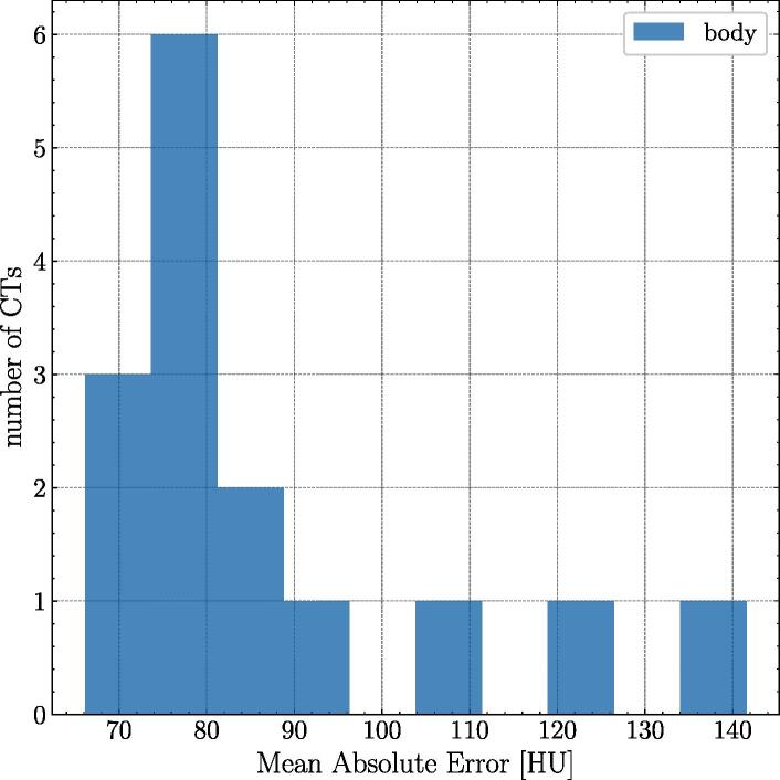
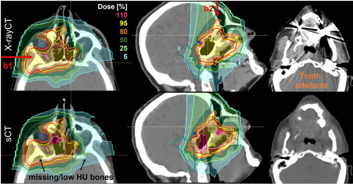
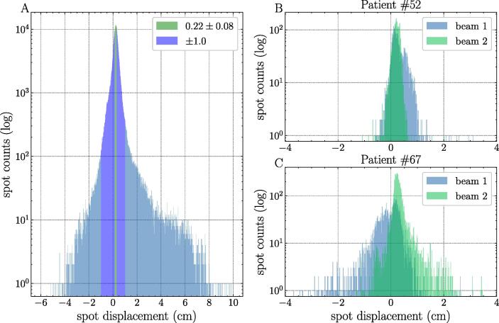
Spot displacement analysis showed a peak displacement by 0.2 cm caused by the immobilisation mask not measurable with the MRI. 95.7% of all spot displacements were located within 1 cm. For the clinical target volume (CTV) the median agreed within −0.2% (−1.3 to 1.4%), while the median differed up to 4.2% (−1.3 to 25.3%) comparing the dose distribution on the X-rayCT and the sCT. OAR deviations depended strongly on the position and the dose gradient. For three patients no deterioration of the OAR parameters was observed. Other patients showed large deteriorations, e.g. for one patient of the chiasm differed by 28.1%.
Conclusion
The usage of sCTs opens several new questions, concluding that we are not ready yet for an MR-only workflow in carbon-ion therapy, as envisaged in photon therapy. Although omitting the X-rayCT seems unfavourable in the case of carbon-ion therapy, an sCT could be advantageous for monitoring, re-planning, and adaptation.
期刊介绍:
Zeitschrift fur Medizinische Physik (Journal of Medical Physics) is an official organ of the German and Austrian Society of Medical Physic and the Swiss Society of Radiobiology and Medical Physics.The Journal is a platform for basic research and practical applications of physical procedures in medical diagnostics and therapy. The articles are reviewed following international standards of peer reviewing.
Focuses of the articles are:
-Biophysical methods in radiation therapy and nuclear medicine
-Dosimetry and radiation protection
-Radiological diagnostics and quality assurance
-Modern imaging techniques, such as computed tomography, magnetic resonance imaging, positron emission tomography
-Ultrasonography diagnostics, application of laser and UV rays
-Electronic processing of biosignals
-Artificial intelligence and machine learning in medical physics
In the Journal, the latest scientific insights find their expression in the form of original articles, reviews, technical communications, and information for the clinical practice.


 分享
分享
 求助内容:
求助内容: 应助结果提醒方式:
 扫码关注我们
