{"title":"腔内动脉传输伪影作为颅内大动脉狭窄的三维飞行时间MR血管造影预测指标:扩大动脉自旋标记MRI在缺血性脑卒中中的应用。","authors":"Sameer Peer, Paramdeep Singh","doi":"10.25259/JCIS_27_2023","DOIUrl":null,"url":null,"abstract":"<p><strong>Objectives: </strong>The objective of this study was to evaluate the diagnostic value of \"intraluminal arterial transit artifact\" in the prediction of intracranial large artery stenosis and to determine if this finding is predictive of ischemic stroke in the territory of the involved artery.</p><p><strong>Material and methods: </strong>The presence of arterial transit artifact (ATA) within the lumen of an intracranial large vessel was noted on three-dimensional time of flight (3D-TOF) magnetic resonance angiography (MRA) (ATA group). The patients with stenosis but with no ATA (no-ATA group), patients with total occlusion (total occlusion group), and patients with no stenosis/occlusion (normal group) were included in the analysis.</p><p><strong>Results: </strong>There were four groups of patients included in the final analysis, the ATA group (<i>n</i> = 22), the no-ATA group (<i>n</i> = 23), the normal group (<i>n</i> = 25), and the total occlusion group (<i>n</i> = 9). Among patients with any demonstrable stenosis (<i>n</i> = 45), the presence of ATA within the stenotic segment was predictive of stenosis of ≥56% (Sensitivity of 100% [85.2-100, 95% CI], specificity of 100% [86.4-100, 95% CI]), with area under curve of 1.0 (0.92-.0, 95% CI). The presence of intra-arterial ATA signal was significantly associated with ischemic stroke as compared with the no-ATA group (86.36% vs. 26.08%, <i>P</i> = 0.0003). Intraluminal ATA was found to be an independent predictor of infarction in the territory of the involved artery.</p><p><strong>Conclusion: </strong>Intraluminal ATA is predictive of stenosis of at least 56% in the involved artery on 3D-TOF MRA. Intraluminal ATA sign may be an independent predictor of infarction in the territory of the involved artery.</p>","PeriodicalId":15512,"journal":{"name":"Journal of Clinical Imaging Science","volume":"13 ","pages":"17"},"PeriodicalIF":1.3000,"publicationDate":"2023-01-01","publicationTypes":"Journal Article","fieldsOfStudy":null,"isOpenAccess":false,"openAccessPdf":"https://ftp.ncbi.nlm.nih.gov/pub/pmc/oa_pdf/11/82/JCIS-13-17.PMC10316254.pdf","citationCount":"0","resultStr":"{\"title\":\"Intraluminal arterial transit artifact as a predictor of intracranial large artery stenosis on 3D time of flight MR angiography: Expanding the application of arterial spin labeling MRI in ischemic stroke.\",\"authors\":\"Sameer Peer, Paramdeep Singh\",\"doi\":\"10.25259/JCIS_27_2023\",\"DOIUrl\":null,\"url\":null,\"abstract\":\"<p><strong>Objectives: </strong>The objective of this study was to evaluate the diagnostic value of \\\"intraluminal arterial transit artifact\\\" in the prediction of intracranial large artery stenosis and to determine if this finding is predictive of ischemic stroke in the territory of the involved artery.</p><p><strong>Material and methods: </strong>The presence of arterial transit artifact (ATA) within the lumen of an intracranial large vessel was noted on three-dimensional time of flight (3D-TOF) magnetic resonance angiography (MRA) (ATA group). The patients with stenosis but with no ATA (no-ATA group), patients with total occlusion (total occlusion group), and patients with no stenosis/occlusion (normal group) were included in the analysis.</p><p><strong>Results: </strong>There were four groups of patients included in the final analysis, the ATA group (<i>n</i> = 22), the no-ATA group (<i>n</i> = 23), the normal group (<i>n</i> = 25), and the total occlusion group (<i>n</i> = 9). Among patients with any demonstrable stenosis (<i>n</i> = 45), the presence of ATA within the stenotic segment was predictive of stenosis of ≥56% (Sensitivity of 100% [85.2-100, 95% CI], specificity of 100% [86.4-100, 95% CI]), with area under curve of 1.0 (0.92-.0, 95% CI). The presence of intra-arterial ATA signal was significantly associated with ischemic stroke as compared with the no-ATA group (86.36% vs. 26.08%, <i>P</i> = 0.0003). Intraluminal ATA was found to be an independent predictor of infarction in the territory of the involved artery.</p><p><strong>Conclusion: </strong>Intraluminal ATA is predictive of stenosis of at least 56% in the involved artery on 3D-TOF MRA. Intraluminal ATA sign may be an independent predictor of infarction in the territory of the involved artery.</p>\",\"PeriodicalId\":15512,\"journal\":{\"name\":\"Journal of Clinical Imaging Science\",\"volume\":\"13 \",\"pages\":\"17\"},\"PeriodicalIF\":1.3000,\"publicationDate\":\"2023-01-01\",\"publicationTypes\":\"Journal Article\",\"fieldsOfStudy\":null,\"isOpenAccess\":false,\"openAccessPdf\":\"https://ftp.ncbi.nlm.nih.gov/pub/pmc/oa_pdf/11/82/JCIS-13-17.PMC10316254.pdf\",\"citationCount\":\"0\",\"resultStr\":null,\"platform\":\"Semanticscholar\",\"paperid\":null,\"PeriodicalName\":\"Journal of Clinical Imaging Science\",\"FirstCategoryId\":\"1085\",\"ListUrlMain\":\"https://doi.org/10.25259/JCIS_27_2023\",\"RegionNum\":0,\"RegionCategory\":null,\"ArticlePicture\":[],\"TitleCN\":null,\"AbstractTextCN\":null,\"PMCID\":null,\"EPubDate\":\"\",\"PubModel\":\"\",\"JCR\":\"Q3\",\"JCRName\":\"RADIOLOGY, NUCLEAR MEDICINE & MEDICAL IMAGING\",\"Score\":null,\"Total\":0}","platform":"Semanticscholar","paperid":null,"PeriodicalName":"Journal of Clinical Imaging Science","FirstCategoryId":"1085","ListUrlMain":"https://doi.org/10.25259/JCIS_27_2023","RegionNum":0,"RegionCategory":null,"ArticlePicture":[],"TitleCN":null,"AbstractTextCN":null,"PMCID":null,"EPubDate":"","PubModel":"","JCR":"Q3","JCRName":"RADIOLOGY, NUCLEAR MEDICINE & MEDICAL IMAGING","Score":null,"Total":0}
Intraluminal arterial transit artifact as a predictor of intracranial large artery stenosis on 3D time of flight MR angiography: Expanding the application of arterial spin labeling MRI in ischemic stroke.
Objectives: The objective of this study was to evaluate the diagnostic value of "intraluminal arterial transit artifact" in the prediction of intracranial large artery stenosis and to determine if this finding is predictive of ischemic stroke in the territory of the involved artery.
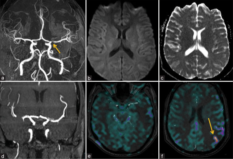
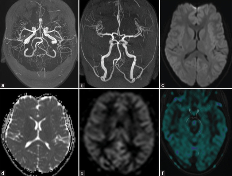
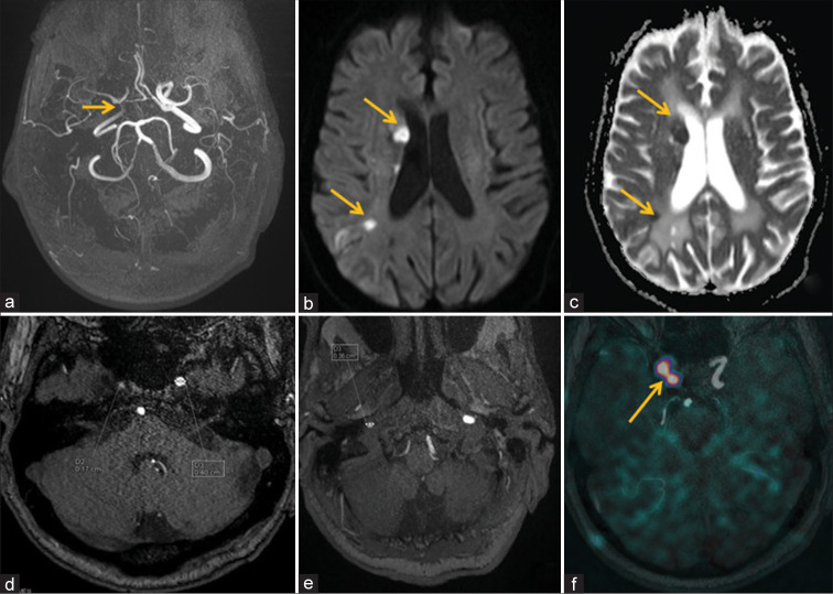
Material and methods: The presence of arterial transit artifact (ATA) within the lumen of an intracranial large vessel was noted on three-dimensional time of flight (3D-TOF) magnetic resonance angiography (MRA) (ATA group). The patients with stenosis but with no ATA (no-ATA group), patients with total occlusion (total occlusion group), and patients with no stenosis/occlusion (normal group) were included in the analysis.
Results: There were four groups of patients included in the final analysis, the ATA group (n = 22), the no-ATA group (n = 23), the normal group (n = 25), and the total occlusion group (n = 9). Among patients with any demonstrable stenosis (n = 45), the presence of ATA within the stenotic segment was predictive of stenosis of ≥56% (Sensitivity of 100% [85.2-100, 95% CI], specificity of 100% [86.4-100, 95% CI]), with area under curve of 1.0 (0.92-.0, 95% CI). The presence of intra-arterial ATA signal was significantly associated with ischemic stroke as compared with the no-ATA group (86.36% vs. 26.08%, P = 0.0003). Intraluminal ATA was found to be an independent predictor of infarction in the territory of the involved artery.
Conclusion: Intraluminal ATA is predictive of stenosis of at least 56% in the involved artery on 3D-TOF MRA. Intraluminal ATA sign may be an independent predictor of infarction in the territory of the involved artery.
期刊介绍:
The Journal of Clinical Imaging Science (JCIS) is an open access peer-reviewed journal committed to publishing high-quality articles in the field of Imaging Science. The journal aims to present Imaging Science and relevant clinical information in an understandable and useful format. The journal is owned and published by the Scientific Scholar. Audience Our audience includes Radiologists, Researchers, Clinicians, medical professionals and students. Review process JCIS has a highly rigorous peer-review process that makes sure that manuscripts are scientifically accurate, relevant, novel and important. Authors disclose all conflicts, affiliations and financial associations such that the published content is not biased.


 分享
分享
 求助内容:
求助内容: 应助结果提醒方式:
 扫码关注我们
