Thore Dietrich, Stephan Theodor Bujak, Thorsten Keller, Bernhard Schnackenburg, Riad Bourayou, Rolf Gebker, Kristof Graf, Eckart Fleck
{"title":"使用1.5特斯拉磁共振成像在体内氟成像描绘实验性心肌炎在啮齿动物模型。","authors":"Thore Dietrich, Stephan Theodor Bujak, Thorsten Keller, Bernhard Schnackenburg, Riad Bourayou, Rolf Gebker, Kristof Graf, Eckart Fleck","doi":"10.1155/2023/4659041","DOIUrl":null,"url":null,"abstract":"<p><p>The usefulness of perfluorocarbon nanoemulsions for the imaging of experimental myocarditis has been demonstrated in a high-field 9.4 Tesla MRI scanner. Our proof-of-concept study investigated the imaging capacity of PFC-based <sup>19</sup>F/<sup>1</sup>H MRI in an animal myocarditis model using a clinical field strength of 1.5 Tesla. To induce experimental myocarditis, five male rats (weight ~300 g, age ~50 days) were treated with one application per week of doxorubicin (2 mg/kg BW) over a period of six weeks. Three control animals received the identical volume of sodium chloride 0.9% instead. Following week six, all animals received a single 4 ml injection of an 20% oil-in-water perfluorooctylbromide nanoemulsion 24 hours prior to <i>in vivo</i><sup>1</sup>H/<sup>19</sup>F imaging on a 1.5 Tesla MRI. After euthanasia, cardiac histology and immunohistochemistry using CD68/ED1 macrophage antibodies were performed, measuring the inflamed myocardium in <i>μ</i>m<sup>2</sup> for further statistical analysis to compare the extent of the inflammation with the <sup>19</sup>F-MRI signal intensity. All animals treated with doxorubicin showed a specific signal in the myocardium, while no myocardial signal could be detected in the control group. Additionally, the doxorubicin group showed a significantly higher SNR for <sup>19</sup>F and a stronger CD68/ED1 immunhistoreactivity compared to the control group. This proof-of-concept study demonstrates that perfluorocarbon nanoemulsions could be detected in an <i>in vivo</i> experimental myocarditis model at a currently clinically relevant field strength.</p>","PeriodicalId":47063,"journal":{"name":"International Journal of Biomedical Imaging","volume":"2023 ","pages":"4659041"},"PeriodicalIF":1.3000,"publicationDate":"2023-01-01","publicationTypes":"Journal Article","fieldsOfStudy":null,"isOpenAccess":false,"openAccessPdf":"https://www.ncbi.nlm.nih.gov/pmc/articles/PMC10361831/pdf/","citationCount":"0","resultStr":"{\"title\":\"In Vivo Fluorine Imaging Using 1.5 Tesla MRI for Depiction of Experimental Myocarditis in a Rodent Animal Model.\",\"authors\":\"Thore Dietrich, Stephan Theodor Bujak, Thorsten Keller, Bernhard Schnackenburg, Riad Bourayou, Rolf Gebker, Kristof Graf, Eckart Fleck\",\"doi\":\"10.1155/2023/4659041\",\"DOIUrl\":null,\"url\":null,\"abstract\":\"<p><p>The usefulness of perfluorocarbon nanoemulsions for the imaging of experimental myocarditis has been demonstrated in a high-field 9.4 Tesla MRI scanner. Our proof-of-concept study investigated the imaging capacity of PFC-based <sup>19</sup>F/<sup>1</sup>H MRI in an animal myocarditis model using a clinical field strength of 1.5 Tesla. To induce experimental myocarditis, five male rats (weight ~300 g, age ~50 days) were treated with one application per week of doxorubicin (2 mg/kg BW) over a period of six weeks. Three control animals received the identical volume of sodium chloride 0.9% instead. Following week six, all animals received a single 4 ml injection of an 20% oil-in-water perfluorooctylbromide nanoemulsion 24 hours prior to <i>in vivo</i><sup>1</sup>H/<sup>19</sup>F imaging on a 1.5 Tesla MRI. After euthanasia, cardiac histology and immunohistochemistry using CD68/ED1 macrophage antibodies were performed, measuring the inflamed myocardium in <i>μ</i>m<sup>2</sup> for further statistical analysis to compare the extent of the inflammation with the <sup>19</sup>F-MRI signal intensity. All animals treated with doxorubicin showed a specific signal in the myocardium, while no myocardial signal could be detected in the control group. Additionally, the doxorubicin group showed a significantly higher SNR for <sup>19</sup>F and a stronger CD68/ED1 immunhistoreactivity compared to the control group. This proof-of-concept study demonstrates that perfluorocarbon nanoemulsions could be detected in an <i>in vivo</i> experimental myocarditis model at a currently clinically relevant field strength.</p>\",\"PeriodicalId\":47063,\"journal\":{\"name\":\"International Journal of Biomedical Imaging\",\"volume\":\"2023 \",\"pages\":\"4659041\"},\"PeriodicalIF\":1.3000,\"publicationDate\":\"2023-01-01\",\"publicationTypes\":\"Journal Article\",\"fieldsOfStudy\":null,\"isOpenAccess\":false,\"openAccessPdf\":\"https://www.ncbi.nlm.nih.gov/pmc/articles/PMC10361831/pdf/\",\"citationCount\":\"0\",\"resultStr\":null,\"platform\":\"Semanticscholar\",\"paperid\":null,\"PeriodicalName\":\"International Journal of Biomedical Imaging\",\"FirstCategoryId\":\"1085\",\"ListUrlMain\":\"https://doi.org/10.1155/2023/4659041\",\"RegionNum\":0,\"RegionCategory\":null,\"ArticlePicture\":[],\"TitleCN\":null,\"AbstractTextCN\":null,\"PMCID\":null,\"EPubDate\":\"\",\"PubModel\":\"\",\"JCR\":\"Q2\",\"JCRName\":\"ENGINEERING, BIOMEDICAL\",\"Score\":null,\"Total\":0}","platform":"Semanticscholar","paperid":null,"PeriodicalName":"International Journal of Biomedical Imaging","FirstCategoryId":"1085","ListUrlMain":"https://doi.org/10.1155/2023/4659041","RegionNum":0,"RegionCategory":null,"ArticlePicture":[],"TitleCN":null,"AbstractTextCN":null,"PMCID":null,"EPubDate":"","PubModel":"","JCR":"Q2","JCRName":"ENGINEERING, BIOMEDICAL","Score":null,"Total":0}
引用次数: 0
摘要
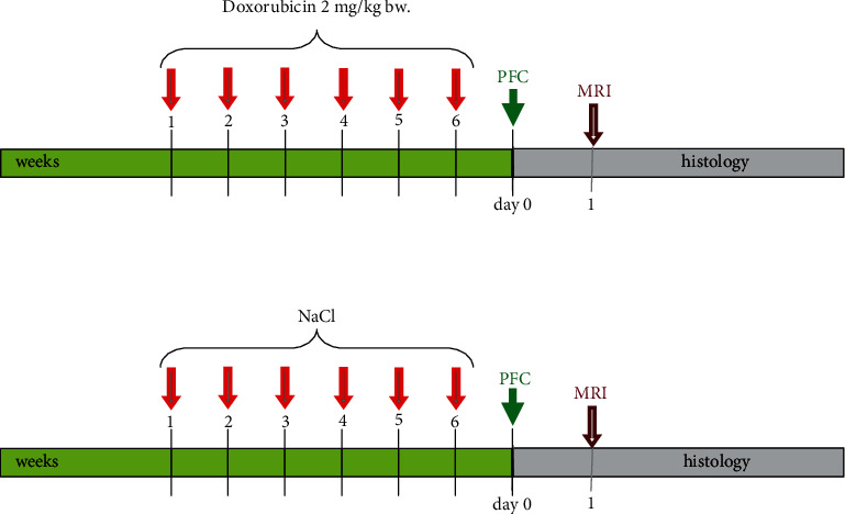
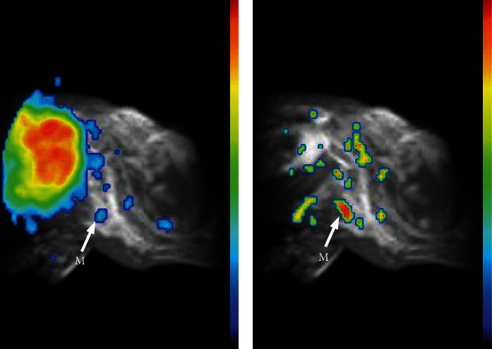
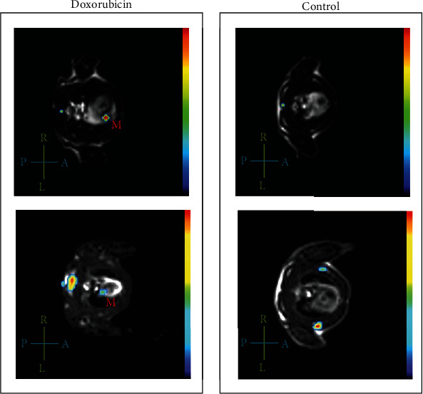
全氟碳纳米乳对实验性心肌炎成像的有用性已在高场9.4特斯拉MRI扫描仪中得到证实。我们的概念验证研究考察了基于pfc的19F/1H MRI在动物心肌炎模型中使用1.5特斯拉临床场强的成像能力。为了诱导实验性心肌炎,将5只体重~300 g、年龄~50日龄的雄性大鼠,每周1次给予阿霉素(2 mg/kg BW),持续6周。而对照组的三只动物则注射了相同体积的0.9%氯化钠。第六周后,所有动物在1.5特斯拉MRI 1h /19F成像前24小时接受单次4 ml 20%水包油全氟辛基溴纳米乳注射。安乐死后采用CD68/ED1巨噬细胞抗体进行心脏组织学和免疫组化,以μm2为单位测量炎症心肌,进一步统计分析炎症程度与19F-MRI信号强度的比较。阿霉素处理的所有动物心肌均有特异信号,而对照组未检测到心肌信号。此外,与对照组相比,阿霉素组显示出更高的19F信噪比和更强的CD68/ED1免疫组化活性。这项概念验证研究表明,全氟碳纳米乳剂可以在体内实验心肌炎模型中以当前临床相关的场强检测到。
In Vivo Fluorine Imaging Using 1.5 Tesla MRI for Depiction of Experimental Myocarditis in a Rodent Animal Model.
The usefulness of perfluorocarbon nanoemulsions for the imaging of experimental myocarditis has been demonstrated in a high-field 9.4 Tesla MRI scanner. Our proof-of-concept study investigated the imaging capacity of PFC-based 19F/1H MRI in an animal myocarditis model using a clinical field strength of 1.5 Tesla. To induce experimental myocarditis, five male rats (weight ~300 g, age ~50 days) were treated with one application per week of doxorubicin (2 mg/kg BW) over a period of six weeks. Three control animals received the identical volume of sodium chloride 0.9% instead. Following week six, all animals received a single 4 ml injection of an 20% oil-in-water perfluorooctylbromide nanoemulsion 24 hours prior to in vivo1H/19F imaging on a 1.5 Tesla MRI. After euthanasia, cardiac histology and immunohistochemistry using CD68/ED1 macrophage antibodies were performed, measuring the inflamed myocardium in μm2 for further statistical analysis to compare the extent of the inflammation with the 19F-MRI signal intensity. All animals treated with doxorubicin showed a specific signal in the myocardium, while no myocardial signal could be detected in the control group. Additionally, the doxorubicin group showed a significantly higher SNR for 19F and a stronger CD68/ED1 immunhistoreactivity compared to the control group. This proof-of-concept study demonstrates that perfluorocarbon nanoemulsions could be detected in an in vivo experimental myocarditis model at a currently clinically relevant field strength.
期刊介绍:
The International Journal of Biomedical Imaging is managed by a board of editors comprising internationally renowned active researchers. The journal is freely accessible online and also offered for purchase in print format. It employs a web-based review system to ensure swift turnaround times while maintaining high standards. In addition to regular issues, special issues are organized by guest editors. The subject areas covered include (but are not limited to):
Digital radiography and tomosynthesis
X-ray computed tomography (CT)
Magnetic resonance imaging (MRI)
Single photon emission computed tomography (SPECT)
Positron emission tomography (PET)
Ultrasound imaging
Diffuse optical tomography, coherence, fluorescence, bioluminescence tomography, impedance tomography
Neutron imaging for biomedical applications
Magnetic and optical spectroscopy, and optical biopsy
Optical, electron, scanning tunneling/atomic force microscopy
Small animal imaging
Functional, cellular, and molecular imaging
Imaging assays for screening and molecular analysis
Microarray image analysis and bioinformatics
Emerging biomedical imaging techniques
Imaging modality fusion
Biomedical imaging instrumentation
Biomedical image processing, pattern recognition, and analysis
Biomedical image visualization, compression, transmission, and storage
Imaging and modeling related to systems biology and systems biomedicine
Applied mathematics, applied physics, and chemistry related to biomedical imaging
Grid-enabling technology for biomedical imaging and informatics


 分享
分享
 求助内容:
求助内容: 应助结果提醒方式:
 扫码关注我们
