{"title":"Endoscopic cadaveric analysis of the origin of the ophthalmic artery.","authors":"Chunhui Zhou, Ting Lei, Junzhao Sun, Hulin Zhao, Xin Yu, Weidong Cao, Wenying Lv, Jianning Zhang","doi":"10.1007/s00276-023-03234-4","DOIUrl":null,"url":null,"abstract":"<p><strong>Purpose: </strong>The ophthalmic artery is often involved in suprasellar and parasellar surgeries, but the anatomical structure where the ophthalmic artery originates has not been fully clarified from the perspective of an endoscopic endonasal approach (EEA).</p><p><strong>Methods: </strong>A total of 10 fresh cadaveric heads (20 sides) were dissected through an EEA, and the origin of the bilateral ophthalmic arteries and their adjacent structures were observed from a ventral view. The origin of the ophthalmic artery in 50 healthy people was retrospectively studied on computed tomography angiography imaging.</p><p><strong>Results: </strong>The ophthalmic artery originated from the intradural segment (75%), paraclinoid segment (15%), or parasellar segment (10%) of the internal carotid artery. The cross-sectional view of the internal carotid artery through the EEA showed that the ophthalmic artery originated from the middle 1/3 (75%) or medial 1/3 (25%) of the upper surface of the internal carotid artery. On computed tomography angiography, the ophthalmic artery originated from the middle 1/3 (77%) and medial 1/3 (22%) of the upper surface of the internal carotid artery. All ophthalmic arteries were near the level of the distal dural ring (DDR) of the internal carotid artery, that is, within 3 mm above or below the DDR.</p><p><strong>Conclusions: </strong>The ophthalmic artery usually originates in the middle 1/3 of the upper surface of the intradural segment of the internal carotid artery within 3 mm of the DDR. The ophthalmic artery can be protected to the utmost extent after its origin is identified through an EEA.</p>","PeriodicalId":49296,"journal":{"name":"Surgical and Radiologic Anatomy","volume":" ","pages":"1435-1441"},"PeriodicalIF":1.2000,"publicationDate":"2023-11-01","publicationTypes":"Journal Article","fieldsOfStudy":null,"isOpenAccess":false,"openAccessPdf":"https://www.ncbi.nlm.nih.gov/pmc/articles/PMC10587203/pdf/","citationCount":"0","resultStr":null,"platform":"Semanticscholar","paperid":null,"PeriodicalName":"Surgical and Radiologic Anatomy","FirstCategoryId":"3","ListUrlMain":"https://doi.org/10.1007/s00276-023-03234-4","RegionNum":4,"RegionCategory":"医学","ArticlePicture":[],"TitleCN":null,"AbstractTextCN":null,"PMCID":null,"EPubDate":"2023/8/18 0:00:00","PubModel":"Epub","JCR":"Q3","JCRName":"ANATOMY & MORPHOLOGY","Score":null,"Total":0}
引用次数: 0
Abstract
Purpose: The ophthalmic artery is often involved in suprasellar and parasellar surgeries, but the anatomical structure where the ophthalmic artery originates has not been fully clarified from the perspective of an endoscopic endonasal approach (EEA).
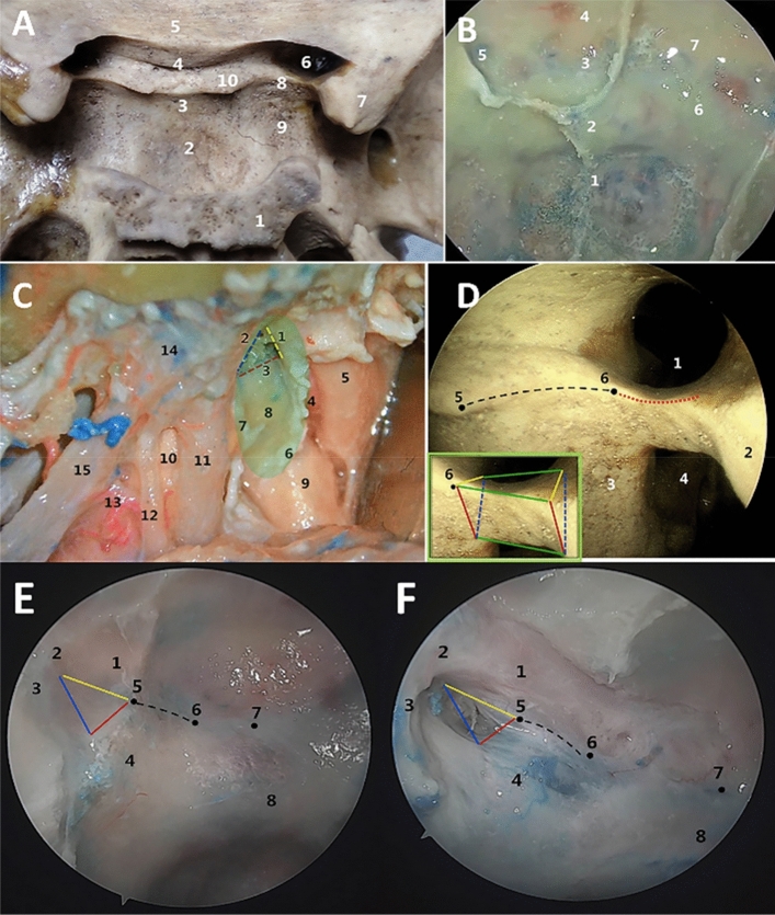
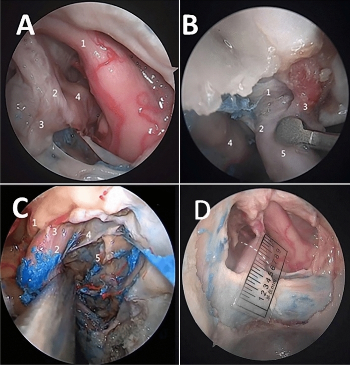
Methods: A total of 10 fresh cadaveric heads (20 sides) were dissected through an EEA, and the origin of the bilateral ophthalmic arteries and their adjacent structures were observed from a ventral view. The origin of the ophthalmic artery in 50 healthy people was retrospectively studied on computed tomography angiography imaging.
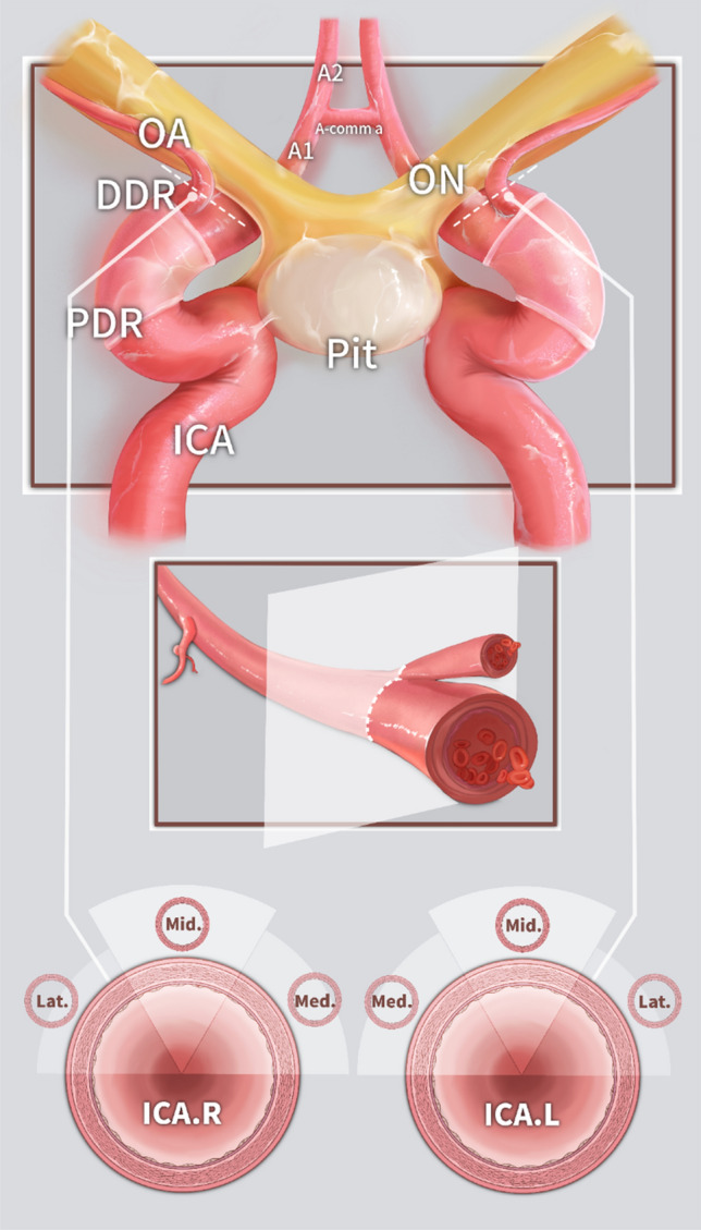
Results: The ophthalmic artery originated from the intradural segment (75%), paraclinoid segment (15%), or parasellar segment (10%) of the internal carotid artery. The cross-sectional view of the internal carotid artery through the EEA showed that the ophthalmic artery originated from the middle 1/3 (75%) or medial 1/3 (25%) of the upper surface of the internal carotid artery. On computed tomography angiography, the ophthalmic artery originated from the middle 1/3 (77%) and medial 1/3 (22%) of the upper surface of the internal carotid artery. All ophthalmic arteries were near the level of the distal dural ring (DDR) of the internal carotid artery, that is, within 3 mm above or below the DDR.
Conclusions: The ophthalmic artery usually originates in the middle 1/3 of the upper surface of the intradural segment of the internal carotid artery within 3 mm of the DDR. The ophthalmic artery can be protected to the utmost extent after its origin is identified through an EEA.
期刊介绍:
Anatomy is a morphological science which cannot fail to interest the clinician. The practical application of anatomical research to clinical problems necessitates special adaptation and selectivity in choosing from numerous international works. Although there is a tendency to believe that meaningful advances in anatomy are unlikely, constant revision is necessary. Surgical and Radiologic Anatomy, the first international journal of Clinical anatomy has been created in this spirit.
Its goal is to serve clinicians, regardless of speciality-physicians, surgeons, radiologists or other specialists-as an indispensable aid with which they can improve their knowledge of anatomy. Each issue includes: Original papers, review articles, articles on the anatomical bases of medical, surgical and radiological techniques, articles of normal radiologic anatomy, brief reviews of anatomical publications of clinical interest.
Particular attention is given to high quality illustrations, which are indispensable for a better understanding of anatomical problems.
Surgical and Radiologic Anatomy is a journal written by anatomists for clinicians with a special interest in anatomy.


 分享
分享
 求助内容:
求助内容: 应助结果提醒方式:
 扫码关注我们
