V3 segment of the right vertebral artery taking an anomalous posterosuperior course and penetrating occipital bone (wall of the jugular foramen) diagnosed by magnetic resonance angiography
{"title":"V3 segment of the right vertebral artery taking an anomalous posterosuperior course and penetrating occipital bone (wall of the jugular foramen) diagnosed by magnetic resonance angiography","authors":"Akira Uchino, Chihiro Suzuki","doi":"10.1007/s00276-024-03478-8","DOIUrl":null,"url":null,"abstract":"<h3 data-test=\"abstract-sub-heading\">Purpose</h3><p>To describe a case of an anomalous posterosuperior course of the V3 segment of the right vertebral artery (VA) that penetrated the occipital bone (wall of the jugular foramen).</p><h3 data-test=\"abstract-sub-heading\">Methods</h3><p>A 33-year-old healthy woman underwent cranial magnetic resonance (MR) imaging and MR angiography from the upper cervical to the intracranial region using a 3-Tesla scanner to screen for asymptomatic brain lesions, including cerebrovascular diseases.</p><h3 data-test=\"abstract-sub-heading\">Results</h3><p>MR angiography showed no pathological arterial lesions such as aneurysms; however, there was an anomalous posterosuperior course of the V3 segment of the right VA. On MR angiographic source images and coronal reformatted images, the right VA was observed to penetrate the occipital bone lateral to the right hypoglossal canal and is located on the inferoposteromedial wall of the right jugular foramen and enter the posterior fossa at a higher level than the foramen magnum.</p><h3 data-test=\"abstract-sub-heading\">Conclusion</h3><p>We present a case in which the right VA showed an anomalous posterosuperior course at the craniovertebral junction. It is extremely rare for a VA to take a higher course. To our knowledge, this is the first report of such a VA variation in the relevant English-language literature. We speculated that the right VA of our patient was formed by the persistence of one more cephalad primitive artery than the first intersegmental artery, not by the persistence of the primitive hypoglossal artery. Careful observation of MR angiographic source is useful and important for identifying the VA penetrating the occipital bone.</p>","PeriodicalId":49296,"journal":{"name":"Surgical and Radiologic Anatomy","volume":"26 1","pages":""},"PeriodicalIF":1.2000,"publicationDate":"2024-09-18","publicationTypes":"Journal Article","fieldsOfStudy":null,"isOpenAccess":false,"openAccessPdf":"","citationCount":"0","resultStr":null,"platform":"Semanticscholar","paperid":null,"PeriodicalName":"Surgical and Radiologic Anatomy","FirstCategoryId":"3","ListUrlMain":"https://doi.org/10.1007/s00276-024-03478-8","RegionNum":4,"RegionCategory":"医学","ArticlePicture":[],"TitleCN":null,"AbstractTextCN":null,"PMCID":null,"EPubDate":"","PubModel":"","JCR":"Q3","JCRName":"ANATOMY & MORPHOLOGY","Score":null,"Total":0}
引用次数: 0
Abstract
Purpose
To describe a case of an anomalous posterosuperior course of the V3 segment of the right vertebral artery (VA) that penetrated the occipital bone (wall of the jugular foramen).
Methods
A 33-year-old healthy woman underwent cranial magnetic resonance (MR) imaging and MR angiography from the upper cervical to the intracranial region using a 3-Tesla scanner to screen for asymptomatic brain lesions, including cerebrovascular diseases.
Results
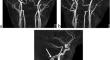
MR angiography showed no pathological arterial lesions such as aneurysms; however, there was an anomalous posterosuperior course of the V3 segment of the right VA. On MR angiographic source images and coronal reformatted images, the right VA was observed to penetrate the occipital bone lateral to the right hypoglossal canal and is located on the inferoposteromedial wall of the right jugular foramen and enter the posterior fossa at a higher level than the foramen magnum.
Conclusion
We present a case in which the right VA showed an anomalous posterosuperior course at the craniovertebral junction. It is extremely rare for a VA to take a higher course. To our knowledge, this is the first report of such a VA variation in the relevant English-language literature. We speculated that the right VA of our patient was formed by the persistence of one more cephalad primitive artery than the first intersegmental artery, not by the persistence of the primitive hypoglossal artery. Careful observation of MR angiographic source is useful and important for identifying the VA penetrating the occipital bone.
目的描述一例右侧椎动脉(VA)V3段异常后上方走向并穿透枕骨(颈静脉孔壁)的病例。方法:一名 33 岁的健康女性使用 3-Tesla 扫描仪接受了头颅磁共振成像(MR)和从上颈部到颅内区域的磁共振血管造影检查,以筛查无症状的脑部病变,包括脑血管疾病。结果磁共振血管造影显示没有动脉瘤等病理性动脉病变,但右侧椎动脉 V3 段的后上方走向异常。在核磁共振血管造影源图像和冠状位重新格式化图像上,观察到右侧 VA 穿透枕骨外侧的右舌下管,位于右侧颈静脉孔内侧壁上,并在比枕骨大孔更高的位置进入后窝。椎间孔走行偏高的情况极为罕见。据我们所知,这是相关英文文献中首次报道这种变异。我们推测患者的右侧 VA 是由于比第一节间动脉多一条头侧原始动脉的持续存在而形成的,而不是原始舌下动脉的持续存在。仔细观察磁共振血管造影的来源对于识别穿透枕骨的 VA 非常有用,也非常重要。
期刊介绍:
Anatomy is a morphological science which cannot fail to interest the clinician. The practical application of anatomical research to clinical problems necessitates special adaptation and selectivity in choosing from numerous international works. Although there is a tendency to believe that meaningful advances in anatomy are unlikely, constant revision is necessary. Surgical and Radiologic Anatomy, the first international journal of Clinical anatomy has been created in this spirit.
Its goal is to serve clinicians, regardless of speciality-physicians, surgeons, radiologists or other specialists-as an indispensable aid with which they can improve their knowledge of anatomy. Each issue includes: Original papers, review articles, articles on the anatomical bases of medical, surgical and radiological techniques, articles of normal radiologic anatomy, brief reviews of anatomical publications of clinical interest.
Particular attention is given to high quality illustrations, which are indispensable for a better understanding of anatomical problems.
Surgical and Radiologic Anatomy is a journal written by anatomists for clinicians with a special interest in anatomy.


 分享
分享
 求助内容:
求助内容: 应助结果提醒方式:
 扫码关注我们
