{"title":"Evaluation and Comparison of Sensitivity and Specificity of Ultrasonography in Placenta Accreta Diagnosis in the Second and Third Trimesters.","authors":"Minoo Movahedi, Farinaz Farahbod, Mahsa Mootamedi","doi":"10.4103/abr.abr_188_22","DOIUrl":null,"url":null,"abstract":"<p><strong>Background: </strong>Ultrasound is the selected technique for the detection of placenta accreta spectrum (PAS). This method can detect PAS in 80%-50% of cases. This study aimed to assess and compare the sensitivity and specificity of ultrasonography in the diagnosis of PAS after the first trimester.</p><p><strong>Materials and methods: </strong>In this prospective study that was performed in 2020-2021 on 79 patients at high risk of PAS, all cases underwent ultrasonography in both 18-22 weeks of gestational age (GA) and 32-34 weeks of GA for evaluation of accreta. As per the risk factors, the delivery plan for all mothers was cesarean section. During the cesarean section, the placenta was examined for accreta, and if it was attached to the uterus, a diagnosis of placenta accreta was ascertained and a total abdominal hysterectomy was performed if the patient's bleeding was not controlled during the operation. The final diagnosis of PAS was made based on the pathology report.</p><p><strong>Results: </strong>Ultrasound evaluation for PAS in 18-22 weeks of GA had 79.17% specificity, 51.61% sensitivity, 61.54% positive predictive value, and 71.70% negative predictive value. Ultrasound imaging for PAS in 32-34 weeks of GA had 60.8% specificity, 90% sensitivity, 62.52% positive predictive value, and 90.33% negative predictive value.</p><p><strong>Conclusion: </strong>It should be concluded that PAS is a critical condition and if the patient is diagnosed in the second or third trimester, special strategies should be applied.</p>","PeriodicalId":7225,"journal":{"name":"Advanced Biomedical Research","volume":"12 ","pages":"188"},"PeriodicalIF":0.0000,"publicationDate":"2023-01-01","publicationTypes":"Journal Article","fieldsOfStudy":null,"isOpenAccess":false,"openAccessPdf":"https://ftp.ncbi.nlm.nih.gov/pub/pmc/oa_pdf/19/06/ABR-12-188.PMC10492602.pdf","citationCount":"0","resultStr":null,"platform":"Semanticscholar","paperid":null,"PeriodicalName":"Advanced Biomedical Research","FirstCategoryId":"1085","ListUrlMain":"https://doi.org/10.4103/abr.abr_188_22","RegionNum":0,"RegionCategory":null,"ArticlePicture":[],"TitleCN":null,"AbstractTextCN":null,"PMCID":null,"EPubDate":"","PubModel":"","JCR":"","JCRName":"","Score":null,"Total":0}
引用次数: 0
Abstract
Background: Ultrasound is the selected technique for the detection of placenta accreta spectrum (PAS). This method can detect PAS in 80%-50% of cases. This study aimed to assess and compare the sensitivity and specificity of ultrasonography in the diagnosis of PAS after the first trimester.
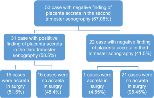
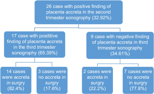
Materials and methods: In this prospective study that was performed in 2020-2021 on 79 patients at high risk of PAS, all cases underwent ultrasonography in both 18-22 weeks of gestational age (GA) and 32-34 weeks of GA for evaluation of accreta. As per the risk factors, the delivery plan for all mothers was cesarean section. During the cesarean section, the placenta was examined for accreta, and if it was attached to the uterus, a diagnosis of placenta accreta was ascertained and a total abdominal hysterectomy was performed if the patient's bleeding was not controlled during the operation. The final diagnosis of PAS was made based on the pathology report.
Results: Ultrasound evaluation for PAS in 18-22 weeks of GA had 79.17% specificity, 51.61% sensitivity, 61.54% positive predictive value, and 71.70% negative predictive value. Ultrasound imaging for PAS in 32-34 weeks of GA had 60.8% specificity, 90% sensitivity, 62.52% positive predictive value, and 90.33% negative predictive value.
Conclusion: It should be concluded that PAS is a critical condition and if the patient is diagnosed in the second or third trimester, special strategies should be applied.


 分享
分享

 求助内容:
求助内容: 应助结果提醒方式:
 扫码关注我们
