{"title":"Connection between medial dorsal cutaneous nerve and saphenous nerve: case report.","authors":"Fatih Çiçek, Turan Koç, Zeliha Kurtoğlu Olgunus","doi":"10.1007/s00276-023-03214-8","DOIUrl":null,"url":null,"abstract":"<p><strong>Purpose: </strong>There are no data on the connection of the saphenous nerve (SN), located on the medial side of the foot, with the terminal branches of the superficial fibular nerve. The aim of this study is to reveal the variation that surgeons should pay attention to for anesthesia applied in foot surgeries.</p><p><strong>Methods: </strong>In this study, the left foot of a 70-year-old female cadaver fixed with formalin was dissected. The distance to the medial malleolus and the incision line was recorded using digital caliper to determine the reference points in the resulting variation.</p><p><strong>Results: </strong>It was observed that a branch from the SN, which arose from the SN and proceeded anteriorly to the upper part of the medial malleolus and continued towards the dorsum of the foot, hooked with a branch from the medial dorsal cutaneous nerve (MDCN). The branches arising from this hook were distributed on the medial edge of the foot up to the proximal metatarsophalangeal joint I. The distance of this nerve connection to the medial malleolus is 91.14 mm, and the distance to the incision line is 15.76 mm.</p><p><strong>Conclusions: </strong>It is suggested that the case presented as an unusual SN variation, which may affect the success of local anesthesia in invasive procedures to the medial part of the foot and could be considered in the evaluation of sensory loss after anteromedial surgical approach to the ankle, should be included in the classification of the cutaneous innervation pattern of the foot.</p>","PeriodicalId":49296,"journal":{"name":"Surgical and Radiologic Anatomy","volume":" ","pages":"1233-1237"},"PeriodicalIF":1.2000,"publicationDate":"2023-10-01","publicationTypes":"Journal Article","fieldsOfStudy":null,"isOpenAccess":false,"openAccessPdf":"","citationCount":"0","resultStr":null,"platform":"Semanticscholar","paperid":null,"PeriodicalName":"Surgical and Radiologic Anatomy","FirstCategoryId":"3","ListUrlMain":"https://doi.org/10.1007/s00276-023-03214-8","RegionNum":4,"RegionCategory":"医学","ArticlePicture":[],"TitleCN":null,"AbstractTextCN":null,"PMCID":null,"EPubDate":"2023/8/1 0:00:00","PubModel":"Epub","JCR":"Q3","JCRName":"ANATOMY & MORPHOLOGY","Score":null,"Total":0}
引用次数: 0
Abstract
Purpose: There are no data on the connection of the saphenous nerve (SN), located on the medial side of the foot, with the terminal branches of the superficial fibular nerve. The aim of this study is to reveal the variation that surgeons should pay attention to for anesthesia applied in foot surgeries.
Methods: In this study, the left foot of a 70-year-old female cadaver fixed with formalin was dissected. The distance to the medial malleolus and the incision line was recorded using digital caliper to determine the reference points in the resulting variation.
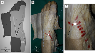
Results: It was observed that a branch from the SN, which arose from the SN and proceeded anteriorly to the upper part of the medial malleolus and continued towards the dorsum of the foot, hooked with a branch from the medial dorsal cutaneous nerve (MDCN). The branches arising from this hook were distributed on the medial edge of the foot up to the proximal metatarsophalangeal joint I. The distance of this nerve connection to the medial malleolus is 91.14 mm, and the distance to the incision line is 15.76 mm.
Conclusions: It is suggested that the case presented as an unusual SN variation, which may affect the success of local anesthesia in invasive procedures to the medial part of the foot and could be considered in the evaluation of sensory loss after anteromedial surgical approach to the ankle, should be included in the classification of the cutaneous innervation pattern of the foot.
期刊介绍:
Anatomy is a morphological science which cannot fail to interest the clinician. The practical application of anatomical research to clinical problems necessitates special adaptation and selectivity in choosing from numerous international works. Although there is a tendency to believe that meaningful advances in anatomy are unlikely, constant revision is necessary. Surgical and Radiologic Anatomy, the first international journal of Clinical anatomy has been created in this spirit.
Its goal is to serve clinicians, regardless of speciality-physicians, surgeons, radiologists or other specialists-as an indispensable aid with which they can improve their knowledge of anatomy. Each issue includes: Original papers, review articles, articles on the anatomical bases of medical, surgical and radiological techniques, articles of normal radiologic anatomy, brief reviews of anatomical publications of clinical interest.
Particular attention is given to high quality illustrations, which are indispensable for a better understanding of anatomical problems.
Surgical and Radiologic Anatomy is a journal written by anatomists for clinicians with a special interest in anatomy.


 分享
分享
 求助内容:
求助内容: 应助结果提醒方式:
 扫码关注我们
