Caveolin-3 loss linked with the P104L LGMD-1C mutation modulates skeletal muscle mTORC1 signalling and cholesterol homeostasis

IF 8.9 1区 医学 Journal of Cachexia, Sarcopenia and Muscle Pub Date : 2023-09-06 DOI:10.1002/jcsm.13317
Dinesh S. Shah, Raid B. Nisr, Gabriela Krasteva-Christ, Harinder S. Hundal
{"title":"Caveolin-3 loss linked with the P104L LGMD-1C mutation modulates skeletal muscle mTORC1 signalling and cholesterol homeostasis","authors":"Dinesh S. Shah,&nbsp;Raid B. Nisr,&nbsp;Gabriela Krasteva-Christ,&nbsp;Harinder S. Hundal","doi":"10.1002/jcsm.13317","DOIUrl":null,"url":null,"abstract":"<div>\n \n \n <section>\n \n <h3> Background</h3>\n \n <p>Caveolins are the principal structural components of plasma membrane caveolae. Dominant pathogenic mutations in the muscle-specific caveolin-3 (Cav3) gene isoform, such as the limb girdle muscular dystrophy type 1C (LGMD-1C) P104L mutation, result in dramatic loss of the Cav3 protein and pathophysiological muscle weakness/wasting. We hypothesize that such muscle degeneration may be linked to disturbances in signalling events that impact protein turnover. Herein, we report studies assessing the effects of Cav3 deficiency on mammalian or mechanistic target of rapamycin complex 1 (mTORC1) signalling in skeletal muscle cells.</p>\n </section>\n \n <section>\n \n <h3> Methods</h3>\n \n <p>L6 myoblasts were stably transfected with Cav3<sup>P104L</sup> or expression of native Cav3 was abolished by CRISPR/Cas9 genome editing (Cav3 knockout [Cav3KO]) prior to performing subcellular fractionation and immunoblotting, analysis of real-time mitochondrial respiration or fixed cell immunocytochemistry. Skeletal muscle from wild-type and Cav3<sup>−/−</sup> mice was processed for immunoblot analysis of downstream mTORC1 substrate phosphorylation.</p>\n </section>\n \n <section>\n \n <h3> Results</h3>\n \n <p>Cav3 was detected in lysosomal-enriched membranes isolated from L6 myoblasts and observed by confocal microscopy to co-localize with lysosomal-specific markers. Cav3<sup>P104L</sup> expression, which results in significant (~95%) loss of native Cav3, or CRISPR/Cas9-mediated Cav3KO, reduced amino acid-dependent mTORC1 activation. The decline in mTORC1-directed signalling was detected by immunoblot analysis of L6 muscle cells and gastrocnemius Cav3<sup>−/−</sup> mouse muscle as judged by reduced phosphorylation of mTORC1 substrates that play key roles in the initiation of protein synthesis (4EBP1<sup>S65</sup> and S6K1<sup>T389</sup>). S6K1<sup>T389</sup> and 4EBP1<sup>S65</sup> phosphorylation reduced by over 75% and 80% in Cav3KO muscle cells and by over 90% and 30% in Cav3<sup>−/−</sup> mouse skeletal muscle, respectively. The reduction in protein synthetic capacity in L6 muscle cells was confirmed by analysis of puromycylated peptides using the SUnSET assay. Cav3 loss was also associated with a 26% increase in lysosomal cholesterol, and pharmacological manipulation of lysosomal cholesterol was effective in replicating the reduction in mTORC1 activity observed in Cav3KO cells. Notably, re-expression of Cav3 in Cav3KO myoblasts normalized lysosomal cholesterol content, which coincided with a recovery in protein translation and an associated increase in mTORC1-directed phosphorylation of downstream targets.</p>\n </section>\n \n <section>\n \n <h3> Conclusions</h3>\n \n <p>Our findings indicate that Cav3 can localize on lysosomal membranes and is a novel regulator of mTORC1 signalling in muscle. Cav3 deficiency associated with the Cav3<sup>P104L</sup> mutation impairs mTORC1 activation and protein synthetic capacity in skeletal muscle cells, which may be linked to disturbances in lysosomal cholesterol trafficking and contribute to the pathology of LGMD-1C.</p>\n </section>\n </div>","PeriodicalId":186,"journal":{"name":"Journal of Cachexia, Sarcopenia and Muscle","volume":"14 5","pages":"2310-2326"},"PeriodicalIF":8.9000,"publicationDate":"2023-09-06","publicationTypes":"Journal Article","fieldsOfStudy":null,"isOpenAccess":false,"openAccessPdf":"https://onlinelibrary.wiley.com/doi/epdf/10.1002/jcsm.13317","citationCount":"0","resultStr":null,"platform":"Semanticscholar","paperid":null,"PeriodicalName":"Journal of Cachexia, Sarcopenia and Muscle","FirstCategoryId":"3","ListUrlMain":"https://onlinelibrary.wiley.com/doi/10.1002/jcsm.13317","RegionNum":1,"RegionCategory":"医学","ArticlePicture":[],"TitleCN":null,"AbstractTextCN":null,"PMCID":null,"EPubDate":"","PubModel":"","JCR":"","JCRName":"","Score":null,"Total":0}
引用次数: 0

Abstract

Background

Caveolins are the principal structural components of plasma membrane caveolae. Dominant pathogenic mutations in the muscle-specific caveolin-3 (Cav3) gene isoform, such as the limb girdle muscular dystrophy type 1C (LGMD-1C) P104L mutation, result in dramatic loss of the Cav3 protein and pathophysiological muscle weakness/wasting. We hypothesize that such muscle degeneration may be linked to disturbances in signalling events that impact protein turnover. Herein, we report studies assessing the effects of Cav3 deficiency on mammalian or mechanistic target of rapamycin complex 1 (mTORC1) signalling in skeletal muscle cells.

Methods

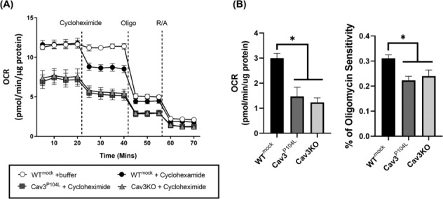
L6 myoblasts were stably transfected with Cav3P104L or expression of native Cav3 was abolished by CRISPR/Cas9 genome editing (Cav3 knockout [Cav3KO]) prior to performing subcellular fractionation and immunoblotting, analysis of real-time mitochondrial respiration or fixed cell immunocytochemistry. Skeletal muscle from wild-type and Cav3−/− mice was processed for immunoblot analysis of downstream mTORC1 substrate phosphorylation.

Results

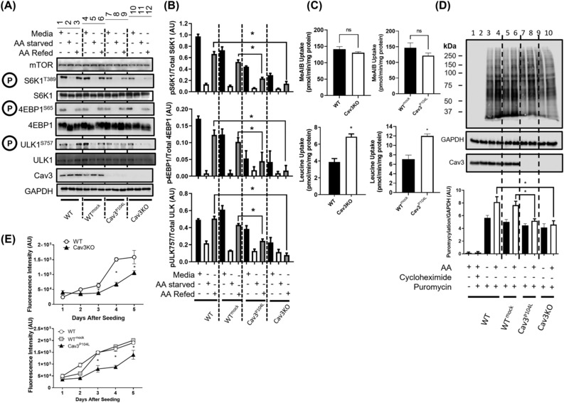
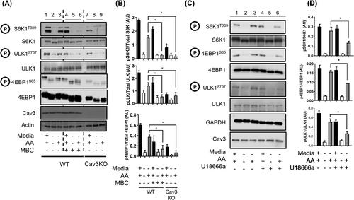
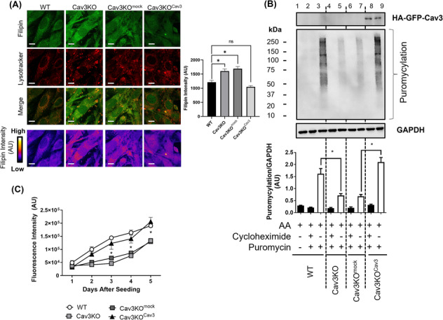
Cav3 was detected in lysosomal-enriched membranes isolated from L6 myoblasts and observed by confocal microscopy to co-localize with lysosomal-specific markers. Cav3P104L expression, which results in significant (~95%) loss of native Cav3, or CRISPR/Cas9-mediated Cav3KO, reduced amino acid-dependent mTORC1 activation. The decline in mTORC1-directed signalling was detected by immunoblot analysis of L6 muscle cells and gastrocnemius Cav3−/− mouse muscle as judged by reduced phosphorylation of mTORC1 substrates that play key roles in the initiation of protein synthesis (4EBP1S65 and S6K1T389). S6K1T389 and 4EBP1S65 phosphorylation reduced by over 75% and 80% in Cav3KO muscle cells and by over 90% and 30% in Cav3−/− mouse skeletal muscle, respectively. The reduction in protein synthetic capacity in L6 muscle cells was confirmed by analysis of puromycylated peptides using the SUnSET assay. Cav3 loss was also associated with a 26% increase in lysosomal cholesterol, and pharmacological manipulation of lysosomal cholesterol was effective in replicating the reduction in mTORC1 activity observed in Cav3KO cells. Notably, re-expression of Cav3 in Cav3KO myoblasts normalized lysosomal cholesterol content, which coincided with a recovery in protein translation and an associated increase in mTORC1-directed phosphorylation of downstream targets.

Conclusions

Our findings indicate that Cav3 can localize on lysosomal membranes and is a novel regulator of mTORC1 signalling in muscle. Cav3 deficiency associated with the Cav3P104L mutation impairs mTORC1 activation and protein synthetic capacity in skeletal muscle cells, which may be linked to disturbances in lysosomal cholesterol trafficking and contribute to the pathology of LGMD-1C.
Abstract Image

Abstract Image

Abstract Image

Abstract Image

查看原文
分享 分享
微信好友 朋友圈 QQ好友 复制链接
本刊更多论文
与P104L LGMD-1C突变相关的Caveolin-3缺失调节骨骼肌mTORC1信号传导和胆固醇稳态。
背景:小窝蛋白是质膜小窝的主要结构成分。肌肉特异性小窝蛋白-3(Cav3)基因亚型的显性致病性突变,如1C型肢带肌营养不良症(LGMD-1C)P104L突变,导致Cav3蛋白的显著损失和病理生理性肌肉无力/萎缩。我们假设这种肌肉退化可能与影响蛋白质周转的信号事件紊乱有关。在此,我们报道了评估Cav3缺乏对骨骼肌细胞中雷帕霉素复合物1(mTORC1)信号传导的哺乳动物或机制靶点的影响的研究。方法:在进行亚细胞分级和免疫印迹、实时线粒体呼吸分析或固定细胞免疫细胞化学之前,用Cav3P104L稳定转染L6成肌细胞,或通过CRISPR/Cas9基因组编辑(Cav3敲除[Cav3KO])消除天然Cav3的表达。对野生型和Cav3-/-小鼠的骨骼肌进行处理,用于下游mTORC1底物磷酸化的免疫印迹分析。结果:Cav3在从L6成肌细胞分离的富含溶酶体的膜中被检测到,并通过共聚焦显微镜观察到与溶酶体特异性标记物共定位。Cav3P104L的表达导致天然Cav3或CRISPR/Cas9介导的Cav3KO的显著(~95%)损失,降低了氨基酸依赖性mTORC1的激活。通过L6肌肉细胞和腓肠肌Cav3-/-小鼠肌肉的免疫印迹分析检测到mTORC1定向信号传导的下降,通过在蛋白质合成起始中起关键作用的mTORC1底物的磷酸化减少来判断(4EBP1S65和S6K1T389)。S6K1T389和4EBP1S65磷酸化在Cav3KO肌肉细胞中分别降低了75%和80%以上,在Cav3-/-小鼠骨骼肌中分别减少了90%和30%以上。L6肌肉细胞中蛋白质合成能力的降低通过使用SUnSET测定的嘌呤酰化肽的分析得到证实。Cav3的损失也与溶酶体胆固醇增加26%有关,溶酶体胆固醇的药理学操作可有效复制在Cav3KO细胞中观察到的mTORC1活性的降低。值得注意的是,Cav3在Cav3KO成肌细胞中的重新表达使溶酶体胆固醇含量正常化,这与蛋白质翻译的恢复和mTORC1引导的下游靶标磷酸化的相关增加相吻合。结论:我们的研究结果表明,Cav3可以定位在溶酶体膜上,是肌肉中mTORC1信号传导的新调节因子。与Cav3P104L突变相关的Cav3缺乏会损害骨骼肌细胞中mTORC1的激活和蛋白质合成能力,这可能与溶酶体胆固醇运输障碍有关,并有助于LGMD-1C的病理学。
本文章由计算机程序翻译,如有差异,请以英文原文为准。
求助全文
约1分钟内获得全文 去求助
来源期刊
Journal of Cachexia, Sarcopenia and Muscle
Journal of Cachexia, Sarcopenia and Muscle Medicine-Orthopedics and Sports Medicine
自引率
12.40%
发文量
0
期刊介绍: The Journal of Cachexia, Sarcopenia, and Muscle is a prestigious, peer-reviewed international publication committed to disseminating research and clinical insights pertaining to cachexia, sarcopenia, body composition, and the physiological and pathophysiological alterations occurring throughout the lifespan and in various illnesses across the spectrum of life sciences. This journal serves as a valuable resource for physicians, biochemists, biologists, dieticians, pharmacologists, and students alike.
期刊最新文献
The Stimulator of Interferon Genes Deficiency Attenuates Diabetic Myopathy Through Inhibiting NLRP3-Mediated Pyroptosis. Assessing Association Between Circulating Bilirubin Levels and the Risk of Frailty: An Observational and Mendelian Randomization Study. Body Composition and Incident High-Intensity Back Pain and/or High Disability: A 10-Year Prospective Population-Based Male Cohort Skeletal Muscle Index Changes on Locoregional Treatment Application After FOLFIRINOX and Survival in Pancreatic Cancer Skeletal Muscle 31P MR Spectroscopy Surpasses CT in Predicting Patient Survival After Liver Transplantation
×
引用
GB/T 7714-2015
复制
MLA
复制
APA
复制
导出至
BibTeX EndNote RefMan NoteFirst NoteExpress
×
×
提示
您的信息不完整,为了账户安全,请先补充。
现在去补充
×
提示
您因"违规操作"
具体请查看互助需知
我知道了
×
提示
现在去查看 取消
×
提示
确定
0
微信
客服QQ
Book学术公众号 扫码关注我们
反馈
×
意见反馈
请填写您的意见或建议
请填写您的手机或邮箱
已复制链接
已复制链接
快去分享给好友吧!
我知道了
×
扫码分享
扫码分享
Book学术官方微信
Book学术文献互助
Book学术文献互助群
群 号:481959085
Book学术
文献互助 智能选刊 最新文献 互助须知 联系我们:info@booksci.cn
Book学术提供免费学术资源搜索服务,方便国内外学者检索中英文文献。致力于提供最便捷和优质的服务体验。
Copyright © 2023 Book学术 All rights reserved.
ghs 京公网安备 11010802042870号 京ICP备2023020795号-1