Correlation between Phase-difference-enhanced MR Imaging and Amyloid Positron Emission Tomography: A Study on Alzheimer's Disease Patients and Normal Controls.
{"title":"Correlation between Phase-difference-enhanced MR Imaging and Amyloid Positron Emission Tomography: A Study on Alzheimer's Disease Patients and Normal Controls.","authors":"Hirotaka Takita, Satoshi Doishita, Tetsuya Yoneda, Hiroyuki Tatekawa, Takato Abe, Yoshiaki Itoh, Daisuke Horiuchi, Taro Tsukamoto, Taro Shimono, Yukio Miki","doi":"10.2463/mrms.mp.2021-0123","DOIUrl":null,"url":null,"abstract":"<p><strong>Purpose: </strong>While amyloid-β deposition in the cerebral cortex for Alzheimer's disease (AD) is often evaluated by amyloid positron emission tomography (PET), amyloid-β-related iron can be detected using phase difference enhanced (PADRE) imaging; however, no study has validated the association between PADRE imaging and amyloid PET. This study investigated whether the degree of hypointense areas on PADRE imaging correlated with the uptake of amyloid PET.</p><p><strong>Methods: </strong>PADRE imaging and amyloid PET were performed in 8 patients with AD and 10 age-matched normal controls. ROIs in the cuneus, precuneus, superior frontal gyrus (SFG), and superior temporal gyrus (STG) were automatically segmented. The degree of hypointense areas on PADRE imaging in each ROI was evaluated using 4-point scaling of visual assessment or volumetric semiquantitative assessment (the percentage of hypointense volume within each ROI). The mean standardized uptake value ratio (SUVR) of amyloid PET in each ROI was also calculated. The Spearman's correlation coefficient between the 4-point scale of PADRE imaging and SUVR of amyloid PET or between the semiquantitative hypointense volume percentage and SUVR in each ROI was evaluated.</p><p><strong>Results: </strong>In the precuneus, a significant positive correlation was identified between the 4-point scale of PADRE imaging and SUVR of amyloid PET (Rs = 0.5; P = 0.034) in all subjects. In the cuneus, a significant positive correlation was identified between the semiquantitative volume percentage of PADRE imaging and SUVR of amyloid PET (Rs = 0.55; P = 0.02) in all subjects.</p><p><strong>Conclusion: </strong>Amyloid-β-enhancing PADRE imaging can be used to predict the SUVR of amyloid PET, especially in the cuneus and precuneus, and may have the potential to be used for diagnosing AD by detecting amyloid deposition.</p>","PeriodicalId":18119,"journal":{"name":"Magnetic Resonance in Medical Sciences","volume":"22 1","pages":"67-78"},"PeriodicalIF":3.2000,"publicationDate":"2023-01-01","publicationTypes":"Journal Article","fieldsOfStudy":null,"isOpenAccess":false,"openAccessPdf":"https://ftp.ncbi.nlm.nih.gov/pub/pmc/oa_pdf/6b/80/mrms-22-67.PMC9849423.pdf","citationCount":"1","resultStr":null,"platform":"Semanticscholar","paperid":null,"PeriodicalName":"Magnetic Resonance in Medical Sciences","FirstCategoryId":"3","ListUrlMain":"https://doi.org/10.2463/mrms.mp.2021-0123","RegionNum":3,"RegionCategory":"医学","ArticlePicture":[],"TitleCN":null,"AbstractTextCN":null,"PMCID":null,"EPubDate":"","PubModel":"","JCR":"Q2","JCRName":"RADIOLOGY, NUCLEAR MEDICINE & MEDICAL IMAGING","Score":null,"Total":0}
引用次数: 1
Abstract
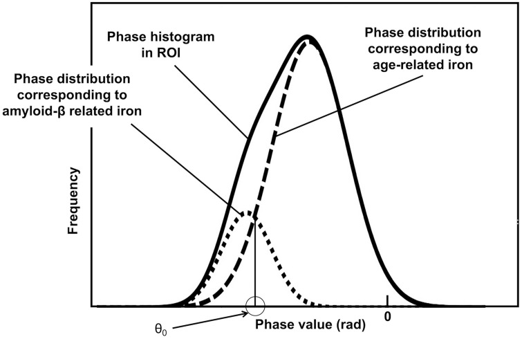
Purpose: While amyloid-β deposition in the cerebral cortex for Alzheimer's disease (AD) is often evaluated by amyloid positron emission tomography (PET), amyloid-β-related iron can be detected using phase difference enhanced (PADRE) imaging; however, no study has validated the association between PADRE imaging and amyloid PET. This study investigated whether the degree of hypointense areas on PADRE imaging correlated with the uptake of amyloid PET.
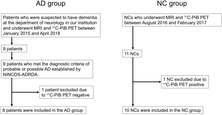
Methods: PADRE imaging and amyloid PET were performed in 8 patients with AD and 10 age-matched normal controls. ROIs in the cuneus, precuneus, superior frontal gyrus (SFG), and superior temporal gyrus (STG) were automatically segmented. The degree of hypointense areas on PADRE imaging in each ROI was evaluated using 4-point scaling of visual assessment or volumetric semiquantitative assessment (the percentage of hypointense volume within each ROI). The mean standardized uptake value ratio (SUVR) of amyloid PET in each ROI was also calculated. The Spearman's correlation coefficient between the 4-point scale of PADRE imaging and SUVR of amyloid PET or between the semiquantitative hypointense volume percentage and SUVR in each ROI was evaluated.
Results: In the precuneus, a significant positive correlation was identified between the 4-point scale of PADRE imaging and SUVR of amyloid PET (Rs = 0.5; P = 0.034) in all subjects. In the cuneus, a significant positive correlation was identified between the semiquantitative volume percentage of PADRE imaging and SUVR of amyloid PET (Rs = 0.55; P = 0.02) in all subjects.
Conclusion: Amyloid-β-enhancing PADRE imaging can be used to predict the SUVR of amyloid PET, especially in the cuneus and precuneus, and may have the potential to be used for diagnosing AD by detecting amyloid deposition.
期刊介绍:
Magnetic Resonance in Medical Sciences (MRMS or Magn
Reson Med Sci) is an international journal pursuing the
publication of original articles contributing to the progress
of magnetic resonance in the field of biomedical sciences
including technical developments and clinical applications.
MRMS is an official journal of the Japanese Society for
Magnetic Resonance in Medicine (JSMRM).


 分享
分享
 求助内容:
求助内容: 应助结果提醒方式:
 扫码关注我们
