Shengbo Gu , Jian Zhuang , Tianying Wang , Shiting Hu , Weilun Song , Xiaobo Liao
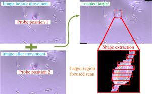
{"title":"The target region focused imaging method for scanning ion conductance microscopy","authors":"Shengbo Gu , Jian Zhuang , Tianying Wang , Shiting Hu , Weilun Song , Xiaobo Liao","doi":"10.1016/j.ultramic.2023.113910","DOIUrl":null,"url":null,"abstract":"<div><p>Scanning ion conductance microscopy (SICM) has developed rapidly and has wide applications in biomedicine, single-cell science and other fields. SICM scanning speed is limited by the conventional raster-type scanning method, which spends most of time on imaging the substrate and does not focus enough on the target area. In order to solve this problem, a target region focused (TRF) method is proposed, which can effectively avoid the scanning of unnecessary substrate areas and enables SICM to image the target area only to achieve high-speed and effective local scanning. TRF method and conventional hopping mode scanning method are compared in the experiments using breast cancer cells and rat basophilic leukemia cells as experimental materials. It was demonstrated that our method can reduce the scanning time for a single sample image significantly without losing scanning information or compromising the quality of imaging. The TRF method developed in this paper can provide an efficient and fast scanning strategy for improving the imaging performance of SICM systems, which can be applied to the dynamic features of cell samples in the fields of biology and pharmacology analysis.</p></div>","PeriodicalId":23439,"journal":{"name":"Ultramicroscopy","volume":"257 ","pages":"Article 113910"},"PeriodicalIF":2.0000,"publicationDate":"2024-03-01","publicationTypes":"Journal Article","fieldsOfStudy":null,"isOpenAccess":false,"openAccessPdf":"","citationCount":"0","resultStr":null,"platform":"Semanticscholar","paperid":null,"PeriodicalName":"Ultramicroscopy","FirstCategoryId":"5","ListUrlMain":"https://www.sciencedirect.com/science/article/pii/S0304399123002279","RegionNum":3,"RegionCategory":"工程技术","ArticlePicture":[],"TitleCN":null,"AbstractTextCN":null,"PMCID":null,"EPubDate":"2023/12/9 0:00:00","PubModel":"Epub","JCR":"Q2","JCRName":"MICROSCOPY","Score":null,"Total":0}
引用次数: 0
Abstract
Scanning ion conductance microscopy (SICM) has developed rapidly and has wide applications in biomedicine, single-cell science and other fields. SICM scanning speed is limited by the conventional raster-type scanning method, which spends most of time on imaging the substrate and does not focus enough on the target area. In order to solve this problem, a target region focused (TRF) method is proposed, which can effectively avoid the scanning of unnecessary substrate areas and enables SICM to image the target area only to achieve high-speed and effective local scanning. TRF method and conventional hopping mode scanning method are compared in the experiments using breast cancer cells and rat basophilic leukemia cells as experimental materials. It was demonstrated that our method can reduce the scanning time for a single sample image significantly without losing scanning information or compromising the quality of imaging. The TRF method developed in this paper can provide an efficient and fast scanning strategy for improving the imaging performance of SICM systems, which can be applied to the dynamic features of cell samples in the fields of biology and pharmacology analysis.
期刊介绍:
Ultramicroscopy is an established journal that provides a forum for the publication of original research papers, invited reviews and rapid communications. The scope of Ultramicroscopy is to describe advances in instrumentation, methods and theory related to all modes of microscopical imaging, diffraction and spectroscopy in the life and physical sciences.


 分享
分享
 求助内容:
求助内容: 应助结果提醒方式:
 扫码关注我们
