Mugurel Constantin Rusu, Răzvan Costin Tudose, Alexandra Diana Vrapciu, Şerban Arghir Popescu
{"title":"Lowered hyoid bone overlapping the thyroid cartilage in CT angiograms","authors":"Mugurel Constantin Rusu, Răzvan Costin Tudose, Alexandra Diana Vrapciu, Şerban Arghir Popescu","doi":"10.1007/s00276-024-03300-5","DOIUrl":null,"url":null,"abstract":"<h3 data-test=\"abstract-sub-heading\">Background</h3><p>The ventral enclosure of the thyroid cartilage by a collapsed hyoid bone (CHB) is poorly encountered in previous research. It was aimed to observe whether or not these malformations could be found and detailed anatomically in a consistent lot of computed tomography (CT) files.</p><h3 data-test=\"abstract-sub-heading\">Methods</h3><p>Two hundred archived CT angiograms were explicitly observed for the CHB anatomical variant.</p><h3 data-test=\"abstract-sub-heading\">Results</h3><p>Different possibilities of CHB were found in 6/200 cases, five males and one female. The symmetrical overlap of the thyroid cartilage by the hyoid body was found in one male case. In three cases, two males and one female, there was asymmetrical overlapping due to tilted hyoid bones. In one male case with such asymmetrical CHD, an ossified anterior longitudinal ligament was noted: the tips of the superior horns of the thyroid cartilage reached lateral to it, thus being retropharyngeal. A different male case had a lowered hyoid with a greater horn fused to the superior horn of the thyroid cartilage, with an interposed ossified triticeal cartilage. In the last male case, the right greater horn collapsed laterally to an ossified triticeal cartilage fused with the thyroid cartilage's superior horn.</p><h3 data-test=\"abstract-sub-heading\">Conclusions</h3><p>The CHB is an undeniable anatomical possibility of an atavism that alters conventional anatomical and surgical landmarks. Different anatomical components of the hyoid bone can descend uni- or bilaterally.</p>","PeriodicalId":49296,"journal":{"name":"Surgical and Radiologic Anatomy","volume":"70 1","pages":""},"PeriodicalIF":1.2000,"publicationDate":"2024-02-05","publicationTypes":"Journal Article","fieldsOfStudy":null,"isOpenAccess":false,"openAccessPdf":"","citationCount":"0","resultStr":null,"platform":"Semanticscholar","paperid":null,"PeriodicalName":"Surgical and Radiologic Anatomy","FirstCategoryId":"3","ListUrlMain":"https://doi.org/10.1007/s00276-024-03300-5","RegionNum":4,"RegionCategory":"医学","ArticlePicture":[],"TitleCN":null,"AbstractTextCN":null,"PMCID":null,"EPubDate":"","PubModel":"","JCR":"Q3","JCRName":"ANATOMY & MORPHOLOGY","Score":null,"Total":0}
引用次数: 0
Abstract
Background
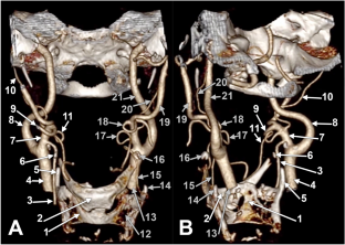
The ventral enclosure of the thyroid cartilage by a collapsed hyoid bone (CHB) is poorly encountered in previous research. It was aimed to observe whether or not these malformations could be found and detailed anatomically in a consistent lot of computed tomography (CT) files.
Methods
Two hundred archived CT angiograms were explicitly observed for the CHB anatomical variant.
Results
Different possibilities of CHB were found in 6/200 cases, five males and one female. The symmetrical overlap of the thyroid cartilage by the hyoid body was found in one male case. In three cases, two males and one female, there was asymmetrical overlapping due to tilted hyoid bones. In one male case with such asymmetrical CHD, an ossified anterior longitudinal ligament was noted: the tips of the superior horns of the thyroid cartilage reached lateral to it, thus being retropharyngeal. A different male case had a lowered hyoid with a greater horn fused to the superior horn of the thyroid cartilage, with an interposed ossified triticeal cartilage. In the last male case, the right greater horn collapsed laterally to an ossified triticeal cartilage fused with the thyroid cartilage's superior horn.
Conclusions
The CHB is an undeniable anatomical possibility of an atavism that alters conventional anatomical and surgical landmarks. Different anatomical components of the hyoid bone can descend uni- or bilaterally.
期刊介绍:
Anatomy is a morphological science which cannot fail to interest the clinician. The practical application of anatomical research to clinical problems necessitates special adaptation and selectivity in choosing from numerous international works. Although there is a tendency to believe that meaningful advances in anatomy are unlikely, constant revision is necessary. Surgical and Radiologic Anatomy, the first international journal of Clinical anatomy has been created in this spirit.
Its goal is to serve clinicians, regardless of speciality-physicians, surgeons, radiologists or other specialists-as an indispensable aid with which they can improve their knowledge of anatomy. Each issue includes: Original papers, review articles, articles on the anatomical bases of medical, surgical and radiological techniques, articles of normal radiologic anatomy, brief reviews of anatomical publications of clinical interest.
Particular attention is given to high quality illustrations, which are indispensable for a better understanding of anatomical problems.
Surgical and Radiologic Anatomy is a journal written by anatomists for clinicians with a special interest in anatomy.


 分享
分享
 求助内容:
求助内容: 应助结果提醒方式:
 扫码关注我们
