James P Higham, Ewan St John Smith, David C Bulmer
{"title":"A note on estimating absolute cytosolic Ca<sup>2+</sup> concentration in sensory neurons using a single wavelength Ca<sup>2+</sup> indicator.","authors":"James P Higham, Ewan St John Smith, David C Bulmer","doi":"10.1177/17448069241230420","DOIUrl":null,"url":null,"abstract":"<p><p>Ca<sup>2+</sup> imaging is frequently used in the investigation of sensory neuronal function and nociception. In vitro imaging of acutely dissociated sensory neurons using membrane-permeant fluorescent Ca<sup>2+</sup> indicators remains the most common approach to study Ca<sup>2+</sup> signalling in sensory neurons. Fluo4 is a popular choice of single-wavelength indicator due to its brightness, high affinity for Ca<sup>2+</sup> and ease of use. However, unlike ratiometric indicators, the emission intensity from single-wavelength indicators can be affected by indicator concentration, optical path length, excitation intensity and detector efficiency. As such, without careful calibration, it can be difficult to draw inferences from differences in the magnitude of Ca<sup>2+</sup> transients recorded using Fluo4. Here, we show that a method scarcely used in sensory neurophysiology - first proposed by Maravall and colleagues (2000) - can provide reliable estimates of absolute cytosolic Ca<sup>2+</sup> concentration ([Ca<sup>2+</sup>]<sub>cyt</sub>) in acutely dissociated sensory neurons using Fluo4. This method is straightforward to implement; is applicable to any high-affinity single-wavelength Ca<sup>2+</sup> indicator with a large dynamic range; and provides estimates of [Ca<sup>2+</sup>]<sub>cyt</sub> in line with other methods, including ratiometric imaging. Use of this method will improve the granularity of sensory neuron Ca<sup>2+</sup> imaging data obtained with Fluo4.</p>","PeriodicalId":19010,"journal":{"name":"Molecular Pain","volume":"20 ","pages":"17448069241230420"},"PeriodicalIF":2.8000,"publicationDate":"2024-01-01","publicationTypes":"Journal Article","fieldsOfStudy":null,"isOpenAccess":false,"openAccessPdf":"https://www.ncbi.nlm.nih.gov/pmc/articles/PMC10880540/pdf/","citationCount":"0","resultStr":null,"platform":"Semanticscholar","paperid":null,"PeriodicalName":"Molecular Pain","FirstCategoryId":"3","ListUrlMain":"https://doi.org/10.1177/17448069241230420","RegionNum":3,"RegionCategory":"医学","ArticlePicture":[],"TitleCN":null,"AbstractTextCN":null,"PMCID":null,"EPubDate":"","PubModel":"","JCR":"Q2","JCRName":"NEUROSCIENCES","Score":null,"Total":0}
引用次数: 0
Abstract
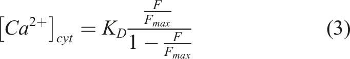
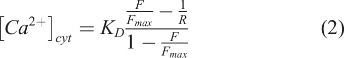
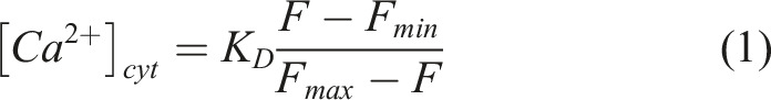
Ca2+ imaging is frequently used in the investigation of sensory neuronal function and nociception. In vitro imaging of acutely dissociated sensory neurons using membrane-permeant fluorescent Ca2+ indicators remains the most common approach to study Ca2+ signalling in sensory neurons. Fluo4 is a popular choice of single-wavelength indicator due to its brightness, high affinity for Ca2+ and ease of use. However, unlike ratiometric indicators, the emission intensity from single-wavelength indicators can be affected by indicator concentration, optical path length, excitation intensity and detector efficiency. As such, without careful calibration, it can be difficult to draw inferences from differences in the magnitude of Ca2+ transients recorded using Fluo4. Here, we show that a method scarcely used in sensory neurophysiology - first proposed by Maravall and colleagues (2000) - can provide reliable estimates of absolute cytosolic Ca2+ concentration ([Ca2+]cyt) in acutely dissociated sensory neurons using Fluo4. This method is straightforward to implement; is applicable to any high-affinity single-wavelength Ca2+ indicator with a large dynamic range; and provides estimates of [Ca2+]cyt in line with other methods, including ratiometric imaging. Use of this method will improve the granularity of sensory neuron Ca2+ imaging data obtained with Fluo4.
期刊介绍:
Molecular Pain is a peer-reviewed, open access journal that considers manuscripts in pain research at the cellular, subcellular and molecular levels. Molecular Pain provides a forum for molecular pain scientists to communicate their research findings in a targeted manner to others in this important and growing field.


 分享
分享
 求助内容:
求助内容: 应助结果提醒方式:
 扫码关注我们
