Maryam Fallahpoor, Dan Nguyen, Ehsan Montahaei, Ali Hosseini, Shahram Nikbakhtian, Maryam Naseri, Faeze Salahshour, Saeed Farzanefar, Mehrshad Abbasi
{"title":"Segmentation of liver and liver lesions using deep learning.","authors":"Maryam Fallahpoor, Dan Nguyen, Ehsan Montahaei, Ali Hosseini, Shahram Nikbakhtian, Maryam Naseri, Faeze Salahshour, Saeed Farzanefar, Mehrshad Abbasi","doi":"10.1007/s13246-024-01390-4","DOIUrl":null,"url":null,"abstract":"<p><p>Segmentation of organs and lesions could be employed for the express purpose of dosimetry in nuclear medicine, assisted image interpretations, and mass image processing studies. Deep leaning created liver and liver lesion segmentation on clinical 3D MRI data has not been fully addressed in previous experiments. To this end, the required data were collected from 128 patients, including their T1w and T2w MRI images, and ground truth labels of the liver and liver lesions were generated. The collection of 110 T1w-T2w MRI image sets was divided, with 94 designated for training and 16 for validation. Furthermore, 18 more datasets were separately allocated for use as hold-out test datasets. The T1w and T2w MRI images were preprocessed into a two-channel format so that they were used as inputs to the deep learning model based on the Isensee 2017 network. To calculate the final Dice coefficient of the network performance on test datasets, the binary average of T1w and T2w predicted images was used. The deep learning model could segment all 18 test cases, with an average Dice coefficient of 88% for the liver and 53% for the liver tumor. Liver segmentation was carried out with rather a high accuracy; this could be achieved for liver dosimetry during systemic or selective radiation therapies as well as for attenuation correction in PET/MRI scanners. Nevertheless, the delineation of liver lesions was not optimal; therefore, tumor detection was not practical by the proposed method on clinical data.</p>","PeriodicalId":48490,"journal":{"name":"Physical and Engineering Sciences in Medicine","volume":" ","pages":"611-619"},"PeriodicalIF":2.0000,"publicationDate":"2024-06-01","publicationTypes":"Journal Article","fieldsOfStudy":null,"isOpenAccess":false,"openAccessPdf":"","citationCount":"0","resultStr":null,"platform":"Semanticscholar","paperid":null,"PeriodicalName":"Physical and Engineering Sciences in Medicine","FirstCategoryId":"3","ListUrlMain":"https://doi.org/10.1007/s13246-024-01390-4","RegionNum":4,"RegionCategory":"医学","ArticlePicture":[],"TitleCN":null,"AbstractTextCN":null,"PMCID":null,"EPubDate":"2024/2/21 0:00:00","PubModel":"Epub","JCR":"Q3","JCRName":"ENGINEERING, BIOMEDICAL","Score":null,"Total":0}
引用次数: 0
Abstract
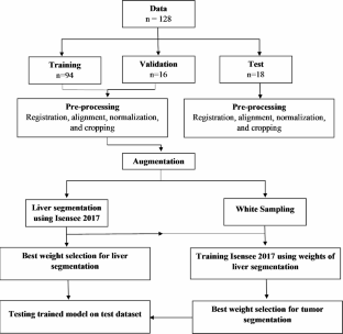
Segmentation of organs and lesions could be employed for the express purpose of dosimetry in nuclear medicine, assisted image interpretations, and mass image processing studies. Deep leaning created liver and liver lesion segmentation on clinical 3D MRI data has not been fully addressed in previous experiments. To this end, the required data were collected from 128 patients, including their T1w and T2w MRI images, and ground truth labels of the liver and liver lesions were generated. The collection of 110 T1w-T2w MRI image sets was divided, with 94 designated for training and 16 for validation. Furthermore, 18 more datasets were separately allocated for use as hold-out test datasets. The T1w and T2w MRI images were preprocessed into a two-channel format so that they were used as inputs to the deep learning model based on the Isensee 2017 network. To calculate the final Dice coefficient of the network performance on test datasets, the binary average of T1w and T2w predicted images was used. The deep learning model could segment all 18 test cases, with an average Dice coefficient of 88% for the liver and 53% for the liver tumor. Liver segmentation was carried out with rather a high accuracy; this could be achieved for liver dosimetry during systemic or selective radiation therapies as well as for attenuation correction in PET/MRI scanners. Nevertheless, the delineation of liver lesions was not optimal; therefore, tumor detection was not practical by the proposed method on clinical data.


 分享
分享
 求助内容:
求助内容: 应助结果提醒方式:
 扫码关注我们
