S Jiménez, I Santos-Álvarez, E Fernández-Valle, D Castejón, P Villa-Valverde, C Rojo-Salvador, P Pérez-Llorens, M. J. Ruiz-Fernández, S. Ariza-Pastrana, R. Martín-Orti, Juncal González-Soriano, Nerea Moreno
{"title":"Comparative MRI analysis of the forebrain of three sauropsida models","authors":"S Jiménez, I Santos-Álvarez, E Fernández-Valle, D Castejón, P Villa-Valverde, C Rojo-Salvador, P Pérez-Llorens, M. J. Ruiz-Fernández, S. Ariza-Pastrana, R. Martín-Orti, Juncal González-Soriano, Nerea Moreno","doi":"10.1007/s00429-024-02788-2","DOIUrl":null,"url":null,"abstract":"<p>The study of the brain by magnetic resonance imaging (MRI) allows to obtain detailed anatomical images, useful to describe specific encephalic structures and to analyze possible variabilities. It is widely used in clinical practice and is becoming increasingly used in veterinary medicine, even in exotic animals; however, despite its potential, its use in comparative neuroanatomy studies is still incipient. It is a technology that in recent years has significantly improved anatomical resolution, together with the fact that it is non-invasive and allows for systematic comparative analysis. All this makes it particularly interesting and useful in evolutionary neuroscience studies, since it allows for the analysis and comparison of brains of rare or otherwise inaccessible species. In the present study, we have analyzed the prosencephalon of three representative sauropsid species, the turtle <i>Trachemys scripta</i> (order Testudine), the lizard <i>Pogona vitticeps</i> (order Squamata) and the snake <i>Python regius</i> (order Squamata) by MRI. In addition, we used MRI sections to analyze the total brain volume and ventricular system of these species, employing volumetric and chemometric analyses together. The raw MRI data of the sauropsida models analyzed in the present study are available for viewing and downloading and have allowed us to produce an atlas of the forebrain of each of the species analyzed, with the main brain regions. In addition, our volumetric data showed that the three groups presented clear differences in terms of total and ventricular brain volumes, particularly the turtles, which in all cases presented distinctive characteristics compared to the lizards and snakes.</p>","PeriodicalId":518000,"journal":{"name":"Brain Structure and Function","volume":"33 1","pages":""},"PeriodicalIF":0.0000,"publicationDate":"2024-03-28","publicationTypes":"Journal Article","fieldsOfStudy":null,"isOpenAccess":false,"openAccessPdf":"","citationCount":"0","resultStr":null,"platform":"Semanticscholar","paperid":null,"PeriodicalName":"Brain Structure and Function","FirstCategoryId":"1085","ListUrlMain":"https://doi.org/10.1007/s00429-024-02788-2","RegionNum":0,"RegionCategory":null,"ArticlePicture":[],"TitleCN":null,"AbstractTextCN":null,"PMCID":null,"EPubDate":"","PubModel":"","JCR":"","JCRName":"","Score":null,"Total":0}
引用次数: 0
Abstract
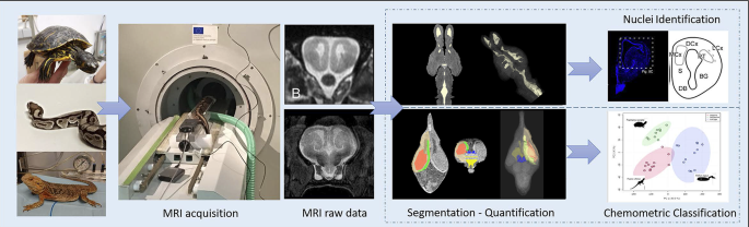
The study of the brain by magnetic resonance imaging (MRI) allows to obtain detailed anatomical images, useful to describe specific encephalic structures and to analyze possible variabilities. It is widely used in clinical practice and is becoming increasingly used in veterinary medicine, even in exotic animals; however, despite its potential, its use in comparative neuroanatomy studies is still incipient. It is a technology that in recent years has significantly improved anatomical resolution, together with the fact that it is non-invasive and allows for systematic comparative analysis. All this makes it particularly interesting and useful in evolutionary neuroscience studies, since it allows for the analysis and comparison of brains of rare or otherwise inaccessible species. In the present study, we have analyzed the prosencephalon of three representative sauropsid species, the turtle Trachemys scripta (order Testudine), the lizard Pogona vitticeps (order Squamata) and the snake Python regius (order Squamata) by MRI. In addition, we used MRI sections to analyze the total brain volume and ventricular system of these species, employing volumetric and chemometric analyses together. The raw MRI data of the sauropsida models analyzed in the present study are available for viewing and downloading and have allowed us to produce an atlas of the forebrain of each of the species analyzed, with the main brain regions. In addition, our volumetric data showed that the three groups presented clear differences in terms of total and ventricular brain volumes, particularly the turtles, which in all cases presented distinctive characteristics compared to the lizards and snakes.


 分享
分享

 求助内容:
求助内容: 应助结果提醒方式:
 扫码关注我们
