{"title":"The calcaneofibular ligament groove at the inferior fibula, an ultrasonographic anatomical landmark","authors":"Akira Kakegawa, Norimi Sumitomo, Ayata Nagira, Yuko Ichinose, Nanae Fukushima","doi":"10.1007/s00276-024-03346-5","DOIUrl":null,"url":null,"abstract":"<h3 data-test=\"abstract-sub-heading\">Purpose</h3><p>Calcaneofibular ligament (CFL) injuries are harder to diagnose than anterior talofibular ligament (ATFL) ones. This study aimed to clarify the fibular attachment of the CFL and verify the bony landmark for evaluating the CFL on ultrasonography.</p><h3 data-test=\"abstract-sub-heading\">Methods</h3><p>Fifty-nine ankles were used in this anatomical study. To confirm the control function of the CFL, we performed passive movement manually using cadaveric ankles and observed the ankle positions where the CFLs were tense.</p><p>Histological observation of CFL attachment of the fibula was performed using Masson’s trichrome stain. The ATFL and CFL were removed, and the bone morphology of the CFL attachment and inferior fibular end was imaged using a stereomicroscope and a 3D scanner. Using ultrasonography, we evaluated the bone morphology of the fibular attachment of the CFL in short-axis images of 27 healthy adult ankles.</p><h3 data-test=\"abstract-sub-heading\">Results</h3><p>The CFL was tensed according to ankle motions: supination, maximum dorsi flexion, maximum plantar flexion, and mild plantar flexion–external rotation. Below the CFL attachment of the fibula was a slight groove between the inferior tip and the obscure tubercle of the fibula. This groove was observed in 81.5% of cases using short-axis ultrasonography.</p><h3 data-test=\"abstract-sub-heading\">Conclusion</h3><p>The CFL was tensed in various ankle positions to control the movements of the talocrural and subtalar joints. There was a slight groove at the inferior end of the fibula where the CFL coursed downward. We called it the CFL groove and proposed that it could serve as a landmark for the short-axis image of ultrasonography.</p>","PeriodicalId":49296,"journal":{"name":"Surgical and Radiologic Anatomy","volume":"5 1","pages":""},"PeriodicalIF":1.2000,"publicationDate":"2024-04-04","publicationTypes":"Journal Article","fieldsOfStudy":null,"isOpenAccess":false,"openAccessPdf":"","citationCount":"0","resultStr":null,"platform":"Semanticscholar","paperid":null,"PeriodicalName":"Surgical and Radiologic Anatomy","FirstCategoryId":"3","ListUrlMain":"https://doi.org/10.1007/s00276-024-03346-5","RegionNum":4,"RegionCategory":"医学","ArticlePicture":[],"TitleCN":null,"AbstractTextCN":null,"PMCID":null,"EPubDate":"","PubModel":"","JCR":"Q3","JCRName":"ANATOMY & MORPHOLOGY","Score":null,"Total":0}
引用次数: 0
Abstract
Purpose
Calcaneofibular ligament (CFL) injuries are harder to diagnose than anterior talofibular ligament (ATFL) ones. This study aimed to clarify the fibular attachment of the CFL and verify the bony landmark for evaluating the CFL on ultrasonography.
Methods
Fifty-nine ankles were used in this anatomical study. To confirm the control function of the CFL, we performed passive movement manually using cadaveric ankles and observed the ankle positions where the CFLs were tense.
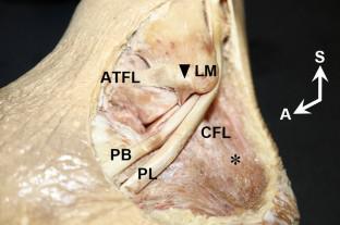
Histological observation of CFL attachment of the fibula was performed using Masson’s trichrome stain. The ATFL and CFL were removed, and the bone morphology of the CFL attachment and inferior fibular end was imaged using a stereomicroscope and a 3D scanner. Using ultrasonography, we evaluated the bone morphology of the fibular attachment of the CFL in short-axis images of 27 healthy adult ankles.
Results
The CFL was tensed according to ankle motions: supination, maximum dorsi flexion, maximum plantar flexion, and mild plantar flexion–external rotation. Below the CFL attachment of the fibula was a slight groove between the inferior tip and the obscure tubercle of the fibula. This groove was observed in 81.5% of cases using short-axis ultrasonography.
Conclusion
The CFL was tensed in various ankle positions to control the movements of the talocrural and subtalar joints. There was a slight groove at the inferior end of the fibula where the CFL coursed downward. We called it the CFL groove and proposed that it could serve as a landmark for the short-axis image of ultrasonography.
期刊介绍:
Anatomy is a morphological science which cannot fail to interest the clinician. The practical application of anatomical research to clinical problems necessitates special adaptation and selectivity in choosing from numerous international works. Although there is a tendency to believe that meaningful advances in anatomy are unlikely, constant revision is necessary. Surgical and Radiologic Anatomy, the first international journal of Clinical anatomy has been created in this spirit.
Its goal is to serve clinicians, regardless of speciality-physicians, surgeons, radiologists or other specialists-as an indispensable aid with which they can improve their knowledge of anatomy. Each issue includes: Original papers, review articles, articles on the anatomical bases of medical, surgical and radiological techniques, articles of normal radiologic anatomy, brief reviews of anatomical publications of clinical interest.
Particular attention is given to high quality illustrations, which are indispensable for a better understanding of anatomical problems.
Surgical and Radiologic Anatomy is a journal written by anatomists for clinicians with a special interest in anatomy.


 分享
分享
 求助内容:
求助内容: 应助结果提醒方式:
 扫码关注我们
