Masuko Ishii, Kota Kato, Koichiro Ichimura, Tatsuo Sakai
{"title":"The three layered structure of orbicularis oris and buccinator complex with partial connection at the modiolus and partial direct continuation","authors":"Masuko Ishii, Kota Kato, Koichiro Ichimura, Tatsuo Sakai","doi":"10.1007/s00276-024-03354-5","DOIUrl":null,"url":null,"abstract":"<h3 data-test=\"abstract-sub-heading\">Purpose</h3><p>This study aimed to clarify the structural arrangement of the orbicularis oris (OOr), the buccinator, and the other perioral muscles around the modiolus.</p><h3 data-test=\"abstract-sub-heading\">Methods</h3><p>The perioral muscles in seventeen cadavers fixed with formalin were dissected in situ and/or in isolated muscle specimens, and their layers were reconstructed schematically upon pantomographic view of the skeleton to evaluate their actions.</p><h3 data-test=\"abstract-sub-heading\">Results</h3><p>The buccinator was composed of three parts including upper and lower oblique parts in its superficial layer and a middle transverse part in its deep layer. The superior and inferior OOr were composed of an inner marginal part (IM) and an outer labial part (OL) in each. The perioral muscles as a whole were arranged in three layers. The first layer consisted of the depressor anguli oris and the OL of superior OOr connected at the modiolus in a vertical direction. The second layer consisted of the upper and inner oblique part of buccinator and a part of the OL of inferior OOr connected at the modiolus in a horizontal direction. The third layer contained the middle transverse part of buccinator continuous with the IM of both OOr and a part of the OL of inferior OOr without connection to the modiolus.</p><h3 data-test=\"abstract-sub-heading\">Conclusions</h3><p>The different arrangement of the three layers of perioral muscles around the modiolus could serve as a good basis to predict the actions of the individual perioral muscles on the movement of lips in open/close of the oral fissure and widening/narrowing of the lip width.</p>","PeriodicalId":49296,"journal":{"name":"Surgical and Radiologic Anatomy","volume":"246 1","pages":""},"PeriodicalIF":1.2000,"publicationDate":"2024-04-04","publicationTypes":"Journal Article","fieldsOfStudy":null,"isOpenAccess":false,"openAccessPdf":"","citationCount":"0","resultStr":null,"platform":"Semanticscholar","paperid":null,"PeriodicalName":"Surgical and Radiologic Anatomy","FirstCategoryId":"3","ListUrlMain":"https://doi.org/10.1007/s00276-024-03354-5","RegionNum":4,"RegionCategory":"医学","ArticlePicture":[],"TitleCN":null,"AbstractTextCN":null,"PMCID":null,"EPubDate":"","PubModel":"","JCR":"Q3","JCRName":"ANATOMY & MORPHOLOGY","Score":null,"Total":0}
引用次数: 0
Abstract
Purpose
This study aimed to clarify the structural arrangement of the orbicularis oris (OOr), the buccinator, and the other perioral muscles around the modiolus.
Methods
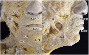
The perioral muscles in seventeen cadavers fixed with formalin were dissected in situ and/or in isolated muscle specimens, and their layers were reconstructed schematically upon pantomographic view of the skeleton to evaluate their actions.
Results
The buccinator was composed of three parts including upper and lower oblique parts in its superficial layer and a middle transverse part in its deep layer. The superior and inferior OOr were composed of an inner marginal part (IM) and an outer labial part (OL) in each. The perioral muscles as a whole were arranged in three layers. The first layer consisted of the depressor anguli oris and the OL of superior OOr connected at the modiolus in a vertical direction. The second layer consisted of the upper and inner oblique part of buccinator and a part of the OL of inferior OOr connected at the modiolus in a horizontal direction. The third layer contained the middle transverse part of buccinator continuous with the IM of both OOr and a part of the OL of inferior OOr without connection to the modiolus.
Conclusions
The different arrangement of the three layers of perioral muscles around the modiolus could serve as a good basis to predict the actions of the individual perioral muscles on the movement of lips in open/close of the oral fissure and widening/narrowing of the lip width.
期刊介绍:
Anatomy is a morphological science which cannot fail to interest the clinician. The practical application of anatomical research to clinical problems necessitates special adaptation and selectivity in choosing from numerous international works. Although there is a tendency to believe that meaningful advances in anatomy are unlikely, constant revision is necessary. Surgical and Radiologic Anatomy, the first international journal of Clinical anatomy has been created in this spirit.
Its goal is to serve clinicians, regardless of speciality-physicians, surgeons, radiologists or other specialists-as an indispensable aid with which they can improve their knowledge of anatomy. Each issue includes: Original papers, review articles, articles on the anatomical bases of medical, surgical and radiological techniques, articles of normal radiologic anatomy, brief reviews of anatomical publications of clinical interest.
Particular attention is given to high quality illustrations, which are indispensable for a better understanding of anatomical problems.
Surgical and Radiologic Anatomy is a journal written by anatomists for clinicians with a special interest in anatomy.


 分享
分享
 求助内容:
求助内容: 应助结果提醒方式:
 扫码关注我们
