{"title":"Intramuscular nerve distribution of the sternocleidomastoid muscle for the botulinum toxin injection","authors":"Melisa Gulcan, Servet Çelik, Canberk Tomruk, Okan Bilge, Yigit Uyanıkgil","doi":"10.1007/s00276-024-03367-0","DOIUrl":null,"url":null,"abstract":"<h3 data-test=\"abstract-sub-heading\">Purpose</h3><p>The aim of this study is to define the intramuscular nerve distribution of the sternocleidomastoid muscle (SCM) and the innervation zones (IZ) to describe the optimal botulinum toxin injection sites.</p><h3 data-test=\"abstract-sub-heading\">Methods</h3><p>The cricoid cartilage (CC), laryngeal prominence (LP) and hyoid bone (HB) and angle of mandible (AM) were determined as landmarks. The length of the muscles were measured between the sternoclavicular joint and tip of the mastoid process. SCM was evaluated in two parts as anterior and posterior divided by the line where the length of the muscle was measured. Measurements were made to define the relationships of the SCM with common carotid artery, internal and external jugular veins. IZ were described according to these vessels. Afterwards, Modified Sihler’s staining technique was applied to expose the intramuscular nerve distribution.</p><h3 data-test=\"abstract-sub-heading\">Results</h3><p>The average length of SCM was 160,1 mm. Motor entry point of the accessory nerve fibers were between the AM-HB lines, in the range of 30–40% of the muscle length, and in the posterior part of the muscles. IZ were between the HB-CC lines in the anterior and posterior part. When this interval was examined according to the vessels, the optimal injection sites were between the LP-CC lines.</p><h3 data-test=\"abstract-sub-heading\">Conclusions</h3><p>This study shows the position of the intramuscular nerve fibers endings of the SCM according to the chosen landmarks and the relationship of the IZ with the vessels to prevent complications. These results can be used as a guide for safe and effective botulinum toxin injections with optimal quantities.</p>","PeriodicalId":49296,"journal":{"name":"Surgical and Radiologic Anatomy","volume":"44 1","pages":""},"PeriodicalIF":1.2000,"publicationDate":"2024-04-29","publicationTypes":"Journal Article","fieldsOfStudy":null,"isOpenAccess":false,"openAccessPdf":"","citationCount":"0","resultStr":null,"platform":"Semanticscholar","paperid":null,"PeriodicalName":"Surgical and Radiologic Anatomy","FirstCategoryId":"3","ListUrlMain":"https://doi.org/10.1007/s00276-024-03367-0","RegionNum":4,"RegionCategory":"医学","ArticlePicture":[],"TitleCN":null,"AbstractTextCN":null,"PMCID":null,"EPubDate":"","PubModel":"","JCR":"Q3","JCRName":"ANATOMY & MORPHOLOGY","Score":null,"Total":0}
引用次数: 0
Abstract
Purpose
The aim of this study is to define the intramuscular nerve distribution of the sternocleidomastoid muscle (SCM) and the innervation zones (IZ) to describe the optimal botulinum toxin injection sites.
Methods
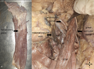
The cricoid cartilage (CC), laryngeal prominence (LP) and hyoid bone (HB) and angle of mandible (AM) were determined as landmarks. The length of the muscles were measured between the sternoclavicular joint and tip of the mastoid process. SCM was evaluated in two parts as anterior and posterior divided by the line where the length of the muscle was measured. Measurements were made to define the relationships of the SCM with common carotid artery, internal and external jugular veins. IZ were described according to these vessels. Afterwards, Modified Sihler’s staining technique was applied to expose the intramuscular nerve distribution.
Results
The average length of SCM was 160,1 mm. Motor entry point of the accessory nerve fibers were between the AM-HB lines, in the range of 30–40% of the muscle length, and in the posterior part of the muscles. IZ were between the HB-CC lines in the anterior and posterior part. When this interval was examined according to the vessels, the optimal injection sites were between the LP-CC lines.
Conclusions
This study shows the position of the intramuscular nerve fibers endings of the SCM according to the chosen landmarks and the relationship of the IZ with the vessels to prevent complications. These results can be used as a guide for safe and effective botulinum toxin injections with optimal quantities.
期刊介绍:
Anatomy is a morphological science which cannot fail to interest the clinician. The practical application of anatomical research to clinical problems necessitates special adaptation and selectivity in choosing from numerous international works. Although there is a tendency to believe that meaningful advances in anatomy are unlikely, constant revision is necessary. Surgical and Radiologic Anatomy, the first international journal of Clinical anatomy has been created in this spirit.
Its goal is to serve clinicians, regardless of speciality-physicians, surgeons, radiologists or other specialists-as an indispensable aid with which they can improve their knowledge of anatomy. Each issue includes: Original papers, review articles, articles on the anatomical bases of medical, surgical and radiological techniques, articles of normal radiologic anatomy, brief reviews of anatomical publications of clinical interest.
Particular attention is given to high quality illustrations, which are indispensable for a better understanding of anatomical problems.
Surgical and Radiologic Anatomy is a journal written by anatomists for clinicians with a special interest in anatomy.


 分享
分享
 求助内容:
求助内容: 应助结果提醒方式:
 扫码关注我们
