Seong-Young Kwon, Sung-Hwan You, Jin Hee Im, Dinh-Huy Nguyen, Dong-Yeon Kim, Ayoung Pyo, Geun-Joong Kim, Hee-Seung Bom, Yeongjin Hong, Jung-Joon Min
{"title":"Tumor Pre-Targeting System Using Streptavidin-Expressing Bacteria.","authors":"Seong-Young Kwon, Sung-Hwan You, Jin Hee Im, Dinh-Huy Nguyen, Dong-Yeon Kim, Ayoung Pyo, Geun-Joong Kim, Hee-Seung Bom, Yeongjin Hong, Jung-Joon Min","doi":"10.1007/s11307-024-01915-z","DOIUrl":null,"url":null,"abstract":"<p><strong>Purpose: </strong>A major obstacle to targeted cancer therapy is identifying suitable targets that are specifically and abundantly expressed by solid tumors. Certain bacterial strains selectively colonize solid tumors and can deliver genetically encoded cargo molecules to the tumor cells. Here, we engineered bacteria to express monomeric streptavidin (mSA) in tumors, and developed a novel tumor pre-targeting system by visualizing the presence of tumor-associated mSA using a biotinylated imaging probe.</p><p><strong>Procedures: </strong>We constructed a plasmid expressing mSA fused to maltose-binding protein and optimized the ribosome binding site sequence to increase solubility and expression levels. E. coli MG1655 was transformed with the recombinant plasmid, expression of which is driven by the pBAD promotor. Expression of mSA was induced by L-arabinose 4 days after injection of bacteria into mice bearing CT26 mouse colon carcinoma cells. Selective accumulation of mSA in tumor tissues was visualized by optical imaging after administration of a biotinylated fluorescent dye. Counting of viable bacterial cells was also performed.</p><p><strong>Results: </strong>Compared with a conventional system, the novel expression system resulted in significantly higher expression of mSA and sustained binding to biotin. Imaging signals in tumor tissues were significantly stronger in the mSA-expressing group than in non-expressing group (P = 0.0005). Furthermore, the fluorescent signal in tumor tissues became detectable again after multiple inductions with L-arabinose. The bacterial counts in tumor tissues showed no significant differences between conditions with and without L-arabinose (P = 0.45). Western blot analysis of tumor tissues confirmed expression and binding of mSA to biotin.</p><p><strong>Conclusions: </strong>We successfully engineered tumor-targeting bacteria carrying a recombinant plasmid expressing mSA, which was targeted to, and expressed in, tumor tissues. These data demonstrate the potential of this novel tumor pre-targeting system when combined with biotinylated imaging probes or therapeutic agents.</p>","PeriodicalId":18760,"journal":{"name":"Molecular Imaging and Biology","volume":" ","pages":"593-602"},"PeriodicalIF":2.5000,"publicationDate":"2024-08-01","publicationTypes":"Journal Article","fieldsOfStudy":null,"isOpenAccess":false,"openAccessPdf":"","citationCount":"0","resultStr":null,"platform":"Semanticscholar","paperid":null,"PeriodicalName":"Molecular Imaging and Biology","FirstCategoryId":"3","ListUrlMain":"https://doi.org/10.1007/s11307-024-01915-z","RegionNum":4,"RegionCategory":"医学","ArticlePicture":[],"TitleCN":null,"AbstractTextCN":null,"PMCID":null,"EPubDate":"2024/5/30 0:00:00","PubModel":"Epub","JCR":"Q2","JCRName":"RADIOLOGY, NUCLEAR MEDICINE & MEDICAL IMAGING","Score":null,"Total":0}
引用次数: 0
Abstract
Purpose: A major obstacle to targeted cancer therapy is identifying suitable targets that are specifically and abundantly expressed by solid tumors. Certain bacterial strains selectively colonize solid tumors and can deliver genetically encoded cargo molecules to the tumor cells. Here, we engineered bacteria to express monomeric streptavidin (mSA) in tumors, and developed a novel tumor pre-targeting system by visualizing the presence of tumor-associated mSA using a biotinylated imaging probe.
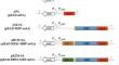
Procedures: We constructed a plasmid expressing mSA fused to maltose-binding protein and optimized the ribosome binding site sequence to increase solubility and expression levels. E. coli MG1655 was transformed with the recombinant plasmid, expression of which is driven by the pBAD promotor. Expression of mSA was induced by L-arabinose 4 days after injection of bacteria into mice bearing CT26 mouse colon carcinoma cells. Selective accumulation of mSA in tumor tissues was visualized by optical imaging after administration of a biotinylated fluorescent dye. Counting of viable bacterial cells was also performed.
Results: Compared with a conventional system, the novel expression system resulted in significantly higher expression of mSA and sustained binding to biotin. Imaging signals in tumor tissues were significantly stronger in the mSA-expressing group than in non-expressing group (P = 0.0005). Furthermore, the fluorescent signal in tumor tissues became detectable again after multiple inductions with L-arabinose. The bacterial counts in tumor tissues showed no significant differences between conditions with and without L-arabinose (P = 0.45). Western blot analysis of tumor tissues confirmed expression and binding of mSA to biotin.
Conclusions: We successfully engineered tumor-targeting bacteria carrying a recombinant plasmid expressing mSA, which was targeted to, and expressed in, tumor tissues. These data demonstrate the potential of this novel tumor pre-targeting system when combined with biotinylated imaging probes or therapeutic agents.
期刊介绍:
Molecular Imaging and Biology (MIB) invites original contributions (research articles, review articles, commentaries, etc.) on the utilization of molecular imaging (i.e., nuclear imaging, optical imaging, autoradiography and pathology, MRI, MPI, ultrasound imaging, radiomics/genomics etc.) to investigate questions related to biology and health. The objective of MIB is to provide a forum to the discovery of molecular mechanisms of disease through the use of imaging techniques. We aim to investigate the biological nature of disease in patients and establish new molecular imaging diagnostic and therapy procedures.
Some areas that are covered are:
Preclinical and clinical imaging of macromolecular targets (e.g., genes, receptors, enzymes) involved in significant biological processes.
The design, characterization, and study of new molecular imaging probes and contrast agents for the functional interrogation of macromolecular targets.
Development and evaluation of imaging systems including instrumentation, image reconstruction algorithms, image analysis, and display.
Development of molecular assay approaches leading to quantification of the biological information obtained in molecular imaging.
Study of in vivo animal models of disease for the development of new molecular diagnostics and therapeutics.
Extension of in vitro and in vivo discoveries using disease models, into well designed clinical research investigations.
Clinical molecular imaging involving clinical investigations, clinical trials and medical management or cost-effectiveness studies.


 分享
分享

 求助内容:
求助内容: 应助结果提醒方式:
 扫码关注我们
