Jagan Mohan, Satish B. Moparthi, Christine Girard-Blanc, Daniele Campisi, Stéphane Blanchard, Charlotte Nugues, Sowmya Rama, Audrey Salles, Esthel Pénard, Stéphane Vassilopoulos, Thomas Wollert
{"title":"ATG16L1 induces the formation of phagophore-like membrane cups","authors":"Jagan Mohan, Satish B. Moparthi, Christine Girard-Blanc, Daniele Campisi, Stéphane Blanchard, Charlotte Nugues, Sowmya Rama, Audrey Salles, Esthel Pénard, Stéphane Vassilopoulos, Thomas Wollert","doi":"10.1038/s41594-024-01300-y","DOIUrl":null,"url":null,"abstract":"The hallmark of non-selective autophagy is the formation of cup-shaped phagophores that capture bulk cytoplasm. The process is accompanied by the conjugation of LC3B to phagophores by an E3 ligase complex comprising ATG12–ATG5 and ATG16L1. Here we combined two complementary reconstitution approaches to reveal the function of LC3B and its ligase complex during phagophore expansion. We found that LC3B forms together with ATG12–ATG5–ATG16L1 a membrane coat that remodels flat membranes into cups that closely resemble phagophores. Mechanistically, we revealed that cup formation strictly depends on a close collaboration between LC3B and ATG16L1. Moreover, only LC3B, but no other member of the ATG8 protein family, promotes cup formation. ATG16L1 truncates that lacked the C-terminal membrane binding domain catalyzed LC3B lipidation but failed to assemble coats, did not promote cup formation and inhibited the biogenesis of non-selective autophagosomes. Our results thus demonstrate that ATG16L1 and LC3B induce and stabilize the characteristic cup-like shape of phagophores. Autophagy degrades cellular waste by engulfing it in phagophore membranes and delivering it to lysosomes for degradation. Here Mohan and colleagues identified a type of membrane coat that assembles on phagophores to guide their expansion.","PeriodicalId":49141,"journal":{"name":"Nature Structural & Molecular Biology","volume":"31 9","pages":"1448-1459"},"PeriodicalIF":10.1000,"publicationDate":"2024-06-04","publicationTypes":"Journal Article","fieldsOfStudy":null,"isOpenAccess":false,"openAccessPdf":"","citationCount":"0","resultStr":null,"platform":"Semanticscholar","paperid":null,"PeriodicalName":"Nature Structural & Molecular Biology","FirstCategoryId":"99","ListUrlMain":"https://www.nature.com/articles/s41594-024-01300-y","RegionNum":1,"RegionCategory":"生物学","ArticlePicture":[],"TitleCN":null,"AbstractTextCN":null,"PMCID":null,"EPubDate":"","PubModel":"","JCR":"Q1","JCRName":"BIOCHEMISTRY & MOLECULAR BIOLOGY","Score":null,"Total":0}
引用次数: 0
Abstract
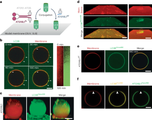
The hallmark of non-selective autophagy is the formation of cup-shaped phagophores that capture bulk cytoplasm. The process is accompanied by the conjugation of LC3B to phagophores by an E3 ligase complex comprising ATG12–ATG5 and ATG16L1. Here we combined two complementary reconstitution approaches to reveal the function of LC3B and its ligase complex during phagophore expansion. We found that LC3B forms together with ATG12–ATG5–ATG16L1 a membrane coat that remodels flat membranes into cups that closely resemble phagophores. Mechanistically, we revealed that cup formation strictly depends on a close collaboration between LC3B and ATG16L1. Moreover, only LC3B, but no other member of the ATG8 protein family, promotes cup formation. ATG16L1 truncates that lacked the C-terminal membrane binding domain catalyzed LC3B lipidation but failed to assemble coats, did not promote cup formation and inhibited the biogenesis of non-selective autophagosomes. Our results thus demonstrate that ATG16L1 and LC3B induce and stabilize the characteristic cup-like shape of phagophores. Autophagy degrades cellular waste by engulfing it in phagophore membranes and delivering it to lysosomes for degradation. Here Mohan and colleagues identified a type of membrane coat that assembles on phagophores to guide their expansion.
期刊介绍:
Nature Structural & Molecular Biology is a comprehensive platform that combines structural and molecular research. Our journal focuses on exploring the functional and mechanistic aspects of biological processes, emphasizing how molecular components collaborate to achieve a particular function. While structural data can shed light on these insights, our publication does not require them as a prerequisite.


 分享
分享
 求助内容:
求助内容: 应助结果提醒方式:
 扫码关注我们
