{"title":"The involvement of Sting in exacerbating acute lung injury in sepsis via the PARP-1/NLRP3 signaling pathway","authors":"Tingting Ying , Yulong Yu , Qimin Yu, Gang Zhou, Lingyang Chen, Yixiao Gu, Lijun Zhu, Haifeng Ying, Minjuan Chen","doi":"10.1016/j.pupt.2024.102303","DOIUrl":null,"url":null,"abstract":"<div><h3>Background</h3><p>Interferon gene stimulator (Sting) is an indispensable adaptor protein that plays a crucial role in acute lung injury (ALI) induced by sepsis, and the PARP-1/NLRP3 signaling pathway may be an integral component of the inflammatory response mediated by Sting. However, the regulatory role of Sting in the PARP-1/NLRP3 pathway in ALI remains insufficiently elucidated.</p></div><div><h3>Methods</h3><p>Using lipopolysaccharide (LPS) to induce ALI in C57BL/6 mice and HUVEC cells, an <em>in vivo</em> and <em>in vitro</em> model was established. <em>In vivo</em>, Sting agonists and inhibitors were administered, while <em>in vitro</em>, Sting was knocked down using siRNA. ELISA was employed to quantify the levels of IL-1β, IL-6, and TNF-α. TUNEL staining was conducted to assess cellular apoptosis, while co-immunoprecipitation was utilized to investigate the interaction between Sting and NLRP3. Expression levels of Sting, NLRP3, PARP-1, among others, were assessed via Western blotting and RT-qPCR. Lung HE staining and lung wet/dry ratio were evaluated in the <em>in vivo</em> mouse model. To validate the role of the PARP-1/NLRP3 signaling pathway, PARP-1 inhibitors were employed both <em>in vivo</em> and <em>in vitro</em>.</p></div><div><h3>Results</h3><p><em>In vitro</em> experiments revealed that the Sting agonist group exacerbated LPS-induced pulmonary pathological damage, pulmonary edema, inflammatory response (increased levels of IL-6, TNF-α, and IL-1β), and cellular injury, whereas the Sting inhibitor group significantly ameliorated the aforementioned injuries, with further improvement observed in the combination therapy of Sting inhibitor and PARP-1 inhibitor. Western blotting and RT-qPCR results demonstrated significant suppression of ICAM-1, VCAM-1, NLRP3, and PARP-1 expression in the Sting inhibitor group, with this reduction further enhanced in the Sting inhibitor + PARP-1 inhibitor treatment group, exhibiting opposite outcomes to the agonist. Furthermore, <em>in vitro</em> experiments using HUVEC cell lines validated these findings.</p></div><div><h3>Conclusions</h3><p>Our study provides new insights into the roles of Sting and the PARP-1/NLRP3 signaling pathway in inflammatory responses, offering novel targets for the development of therapeutic interventions against inflammatory reactions.</p></div>","PeriodicalId":20799,"journal":{"name":"Pulmonary pharmacology & therapeutics","volume":"86 ","pages":"Article 102303"},"PeriodicalIF":3.3000,"publicationDate":"2024-06-05","publicationTypes":"Journal Article","fieldsOfStudy":null,"isOpenAccess":false,"openAccessPdf":"","citationCount":"0","resultStr":null,"platform":"Semanticscholar","paperid":null,"PeriodicalName":"Pulmonary pharmacology & therapeutics","FirstCategoryId":"3","ListUrlMain":"https://www.sciencedirect.com/science/article/pii/S1094553924000191","RegionNum":3,"RegionCategory":"医学","ArticlePicture":[],"TitleCN":null,"AbstractTextCN":null,"PMCID":null,"EPubDate":"","PubModel":"","JCR":"Q2","JCRName":"PHARMACOLOGY & PHARMACY","Score":null,"Total":0}
引用次数: 0
Abstract
Background
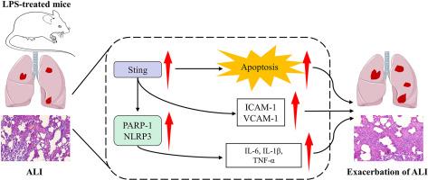
Interferon gene stimulator (Sting) is an indispensable adaptor protein that plays a crucial role in acute lung injury (ALI) induced by sepsis, and the PARP-1/NLRP3 signaling pathway may be an integral component of the inflammatory response mediated by Sting. However, the regulatory role of Sting in the PARP-1/NLRP3 pathway in ALI remains insufficiently elucidated.
Methods
Using lipopolysaccharide (LPS) to induce ALI in C57BL/6 mice and HUVEC cells, an in vivo and in vitro model was established. In vivo, Sting agonists and inhibitors were administered, while in vitro, Sting was knocked down using siRNA. ELISA was employed to quantify the levels of IL-1β, IL-6, and TNF-α. TUNEL staining was conducted to assess cellular apoptosis, while co-immunoprecipitation was utilized to investigate the interaction between Sting and NLRP3. Expression levels of Sting, NLRP3, PARP-1, among others, were assessed via Western blotting and RT-qPCR. Lung HE staining and lung wet/dry ratio were evaluated in the in vivo mouse model. To validate the role of the PARP-1/NLRP3 signaling pathway, PARP-1 inhibitors were employed both in vivo and in vitro.
Results
In vitro experiments revealed that the Sting agonist group exacerbated LPS-induced pulmonary pathological damage, pulmonary edema, inflammatory response (increased levels of IL-6, TNF-α, and IL-1β), and cellular injury, whereas the Sting inhibitor group significantly ameliorated the aforementioned injuries, with further improvement observed in the combination therapy of Sting inhibitor and PARP-1 inhibitor. Western blotting and RT-qPCR results demonstrated significant suppression of ICAM-1, VCAM-1, NLRP3, and PARP-1 expression in the Sting inhibitor group, with this reduction further enhanced in the Sting inhibitor + PARP-1 inhibitor treatment group, exhibiting opposite outcomes to the agonist. Furthermore, in vitro experiments using HUVEC cell lines validated these findings.
Conclusions
Our study provides new insights into the roles of Sting and the PARP-1/NLRP3 signaling pathway in inflammatory responses, offering novel targets for the development of therapeutic interventions against inflammatory reactions.
期刊介绍:
Pulmonary Pharmacology and Therapeutics (formerly Pulmonary Pharmacology) is concerned with lung pharmacology from molecular to clinical aspects. The subject matter encompasses the major diseases of the lung including asthma, cystic fibrosis, pulmonary circulation, ARDS, carcinoma, bronchitis, emphysema and drug delivery. Laboratory and clinical research on man and animals will be considered including studies related to chemotherapy of cancer, tuberculosis and infection. In addition to original research papers the journal will include review articles and book reviews.
Research Areas Include:
• All major diseases of the lung
• Physiology
• Pathology
• Drug delivery
• Metabolism
• Pulmonary Toxicology.


 分享
分享
 求助内容:
求助内容: 应助结果提醒方式:
 扫码关注我们
